The Power of Process: Document Your Journey of Pathology Improvement Through Art
By Anastazia Hartman | October 27The Department of Pathology is excited to announce the winners of our annual department art competition, “The Power of Process: Document Your Journey of Pathology Improvement Through Art”. The competition celebrates the creativity and artistry inspired by the work we do every day. This year’s installation is dedicated to the memory of Regina Ferguson, in recognition of her support and the important role she played in bringing this program and its artwork to life.
--
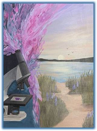 First Place: “A Path of Light” by Lauren Branson, Administrative Specialist
Artist Statement: Through the microscope, we see the complexity and beauty of human tissue, but the Michigan shoreline reminds us to pause and find beauty beyond our work and difficult diagnoses. This piece serves as a reminder that even in our most challenging work, compassion, hope and beauty continue to guide us. Created in honor of Regina Ferguson, who brought so much light to our department.
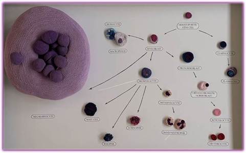 Second Place: “Hematocrochesis” by Camille Van Neste, MD, PhD, Pathology Resident
Artist Statement: This project began with the idea of crocheting nesting cells, and the hematopoietic system is second to none in having morphologically distinct cells of multiple lineages. As the hematopoietic stem cell divides, its progeny trade the potential to become any blood cell for becoming a mature blood cell. During the journey from stem cell to erythrocyte (red blood cell), it must first commit to the myeloid lineage, losing the option of becoming a lymphoid cell. Again, it must be shepherded away from the granulocyte and megakaryocyte pathways to differentiate into the normoblast lineage, eventually losing the ability to divide completely. As an orthochromic normoblast, this cell makes the ultimate sacrifice - it jettisons its nucleus, the genetic code that arguably defined it as a cell at all. Its fate is sealed and its days are numbered - it will spend the next three months circulating the body before it disintegrates and its components are recycled. One can mourn the doors closed and opportunities lost along the way. However, mature erythrocytes are exquisitely adapted to their job: they travel hundreds of miles delivering oxygen to keep every single other cell alive.
Like many doctors, I too took a metaphorical journey of differentiation. At the beginning of medical school, I was undifferentiated and interested in many different medical specialties. However, bit by bit throughout medical school and my PhD, I had a number of small realizations that culminated in a clear choice: I am meant to be a pathologist.
I began hematocrochesis in late 2023, shortly after completing my away rotation at Michigan, and I have been slowly working on it since. I passed a point of no return in my career when I matched into this pathology residency program. The cost of closing doors on the other specialties pales in comparison to the joy I have found in the U-M pathology residency program, where I am able to learn so much in a positive environment and make friends I will hold dear for the rest of my life. In parallel, through roughly 100 hours of crocheting, my understanding of the nuances and blood cell morphology has matured. Soon I will enter fellowship and differentiate further - as I learn, my career focuses on a narrower path but also unlocks new skills and opportunities.
Certain astute viewers will notice that the relative sizes of these cells are not to scale. I designed the cells to be nested - the neutrophil holds the band, which can contain the metamyelocyte, which can contain the myelocyte, etc. Each set of nested cells fits into a megakaryocyte nucleus. The whole system, despite its diversity of cells and level of specialization, all fits together just as we take our past selves at every step of our journey with us towards our more differentiated futures.
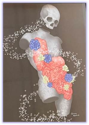 Honorable Mention: “Life in Death” by Olivia Harkins, Pathologist Assistant
Artist Statement: I enjoy exploring my two passions of science and art within my work. “Life in Death” illustrates the permanence of a stone-like figure with more than just plant overgrowth creeping onto the surface; it incorporates anatomy to reflect that, like a post-mortem autopsy, while maintaining the beauty of the human form inside and out.
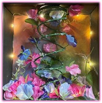 Honorable Mention: “My Time Will Come” by Kimberly Van Dine, Medical Laboratory Scientist
Artist Statement: A sculpture of how the pea plant led to the discovery of DNA and therefore the study of genetics. The pea plant was a model plant because of its easily distinguishable characteristics and true-breeding nature. Gregor Mendel used the pea plant to study how these characteristics could be passed down during the 1850s. This led to the Laws of Segregation and Independent Assortment. While Mendell's findings were mostly ignored during his time, he told his friend Gustav von Niessl "My time will come." In the early 1900s, Hugo de Vries, Carl Corren, Wiliam Jasper Spillman would all independently validate Mendell's findings finally leading to the recognition of Mendel's work in the field of genetics.
This sculpture shows the hidden potential that can be found in an ordinary plant. While we may not be able to see our own significance in what we do here every day, it is still a vital role that can one day help lead to significant milestones.
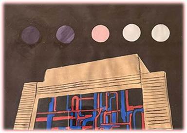 Honorable Mention: "Regina Ferguson" by Cooper Cooper, Histotechnologist
Artist Statement: The H&E stain is the gold standard for medical diagnosis in histology. The stain was introduced in1877 by chemist Nicolaus Wissozky. (Photo credit to the University of Michigan Medical School)
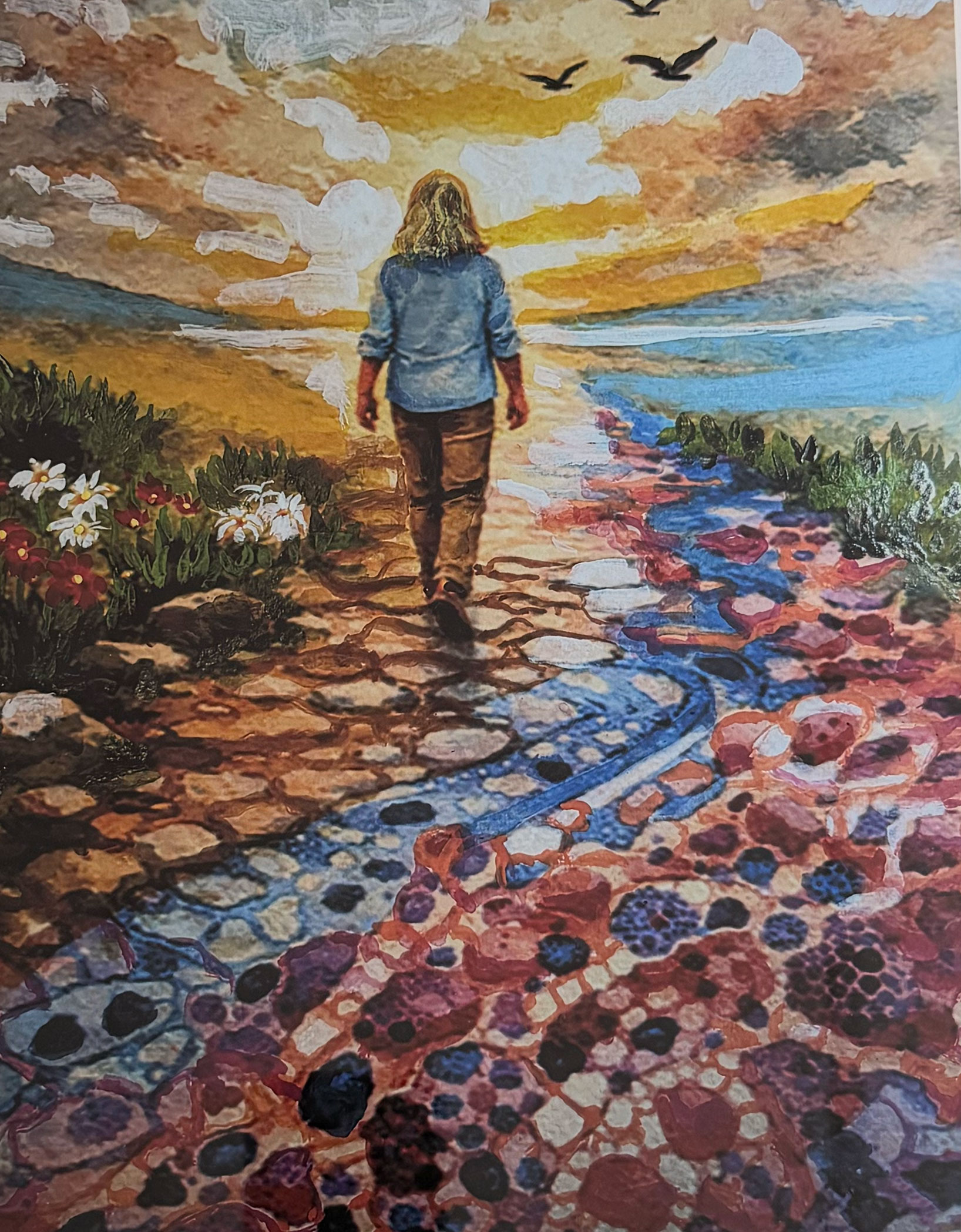 Non-Pathology Participant Recognition: "The Answer Was Always There", Michele Mitchell, Patient Advocate
Artist Statement: Michele Mitchell hopes to highlight through this watercolor that, as a patient, the moment of diagnosis can feel like standing at the edge of an unknown path-uncertain, fragmented, and filled with questions that the body itself cannot fully answer. This watercolor reflects that journey, not as one defined by disease, but by understanding.
In the painting, Michele walks forward toward light and open sky. The path beneath her feet begins as earth-organic, familiar, and gradually transforms into a digital pathology image. What was once invisible becomes foundational. The diagnosis does not stop her movement; it enables it. The answers were always there, waiting to be seen, interpreted, and translated.
Digital pathology represents more than technology-it represents clarity. It is the bridge between uncertainty and understanding, between fear and informed action. The cellular patterns woven into the path are intentionally stylized, not clinical, to reflect how science and humanity coexist. These images are not just data points; they are stories-stories that, when interpreted by pathologists, give patients language, direction, and hope.
"As a patient advocate and cancer survivor, I have learned that thriving begins when patients are invited into the conversation. When pathology is transparent and accessible, it becomes empowering. The path forward becomes visible. This piece honors pathologists as navigators-those who illuminate the unseen and transform microscopic detail into life-changing understanding. The painting reminds us that diagnosis, when guided by precision and compassion, does not define a person. It supports them as they continue forward-grounded, informed, and moving toward possibility."
--
Congratulations to all our winners and honorees! We sincerely thank everyone who participated and shared their artistic talents with our department. Your contributions celebrate the intersection of art, science, and the many stories that make pathology meaningful.
 ON THE COVER
 ON THE COVER
 ON THE COVER
 ON THE COVER
 ON THE COVER
 ON THE COVER
 ON THE COVER
 ON THE COVER
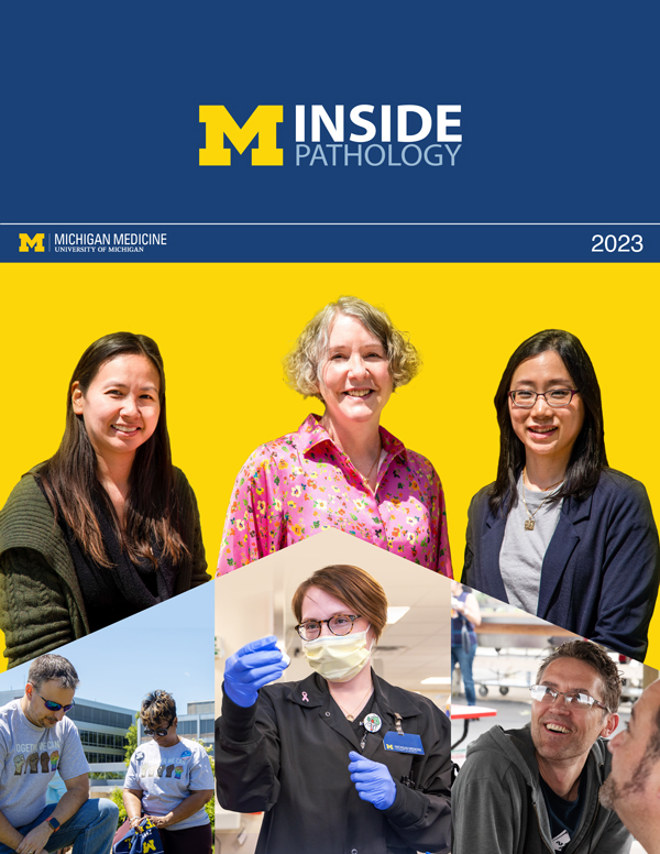 ON THE COVER
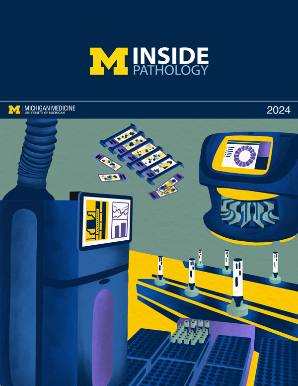 ON THE COVER
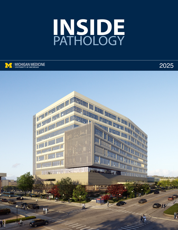 ON THE COVER
