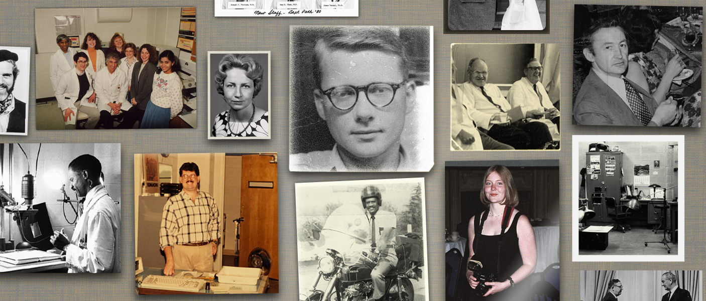
Standing inside the thoroughly modern Pathology Imaging Facility, one glimpses a rich history in a series of photographs preserved behind a glass frame. Most of the images are black and white, some stained and sepia toned with curled edges, evoking an old family album. In one picture, A. James French, M.D., a former department chair, shakes hands with President Lyndon Johnson. In another, Bruce Friedman, M.D., now a professor emeritus, sits in a military jeep in Korea, where he was assigned during the Korean War. One image captures Pathology residents streaking across the Diag. Another shows Kathy Heidelberger Davenport, M.D., now a professor emerita, advising actor Robert Young for the 1970s television series Marcus Welby, M.D. There’s also an image of Paul Gikas, MD, who died peacefully last year after retiring in 1994. In this picture, he’s carving a turkey for a Department Thanksgiving.
Based in the Medical Science 1 Building, Pathology Imaging provides image- and video-making services to the Department. Full-time image specialists assist with everything from photographing autopsies to photographing departmental events. The end products are used for clinical, research, teaching, and promotional purposes. Following the addition of the Wayne County Medical Examiner’s Office (WCMEO) to the Department, Pathology Imaging also employs an imaging specialist at WCMEO to document autopsies and process photo requests for local police departments and the prosecutor’s office.
The Department hired its first full-time photographer, Matt Hardeen, in 1958, at a time when pathology, like radiology, was becoming highly image-driven. There was a growing need for photographs to provide clinical documentation and showcase research results. Before Hardeen’s retirement, J. “Craig” Biddle was hired in 1968, and Eddie Burks joined as a full-time photographer in 1971. Today, there are three full-time image specialists: Mark Deming and Elizabeth Walker at the Pathology Imaging Facility in Ann Arbor, and Kelly Root at WCMEO.
Working for Pathology imaging since 1987, Mark Deming initially served as an assistant for Biddle and Burks. He says the lab was less crowded then, and the darkroom’s photographic print-washing tub was always filled with floating photos. Space was available to hang backdrops for portraits, but as new faculty joined the Department, the lab filled with microscopes and other scientific equipment. Increasingly, faculty members needed clinical images that could provide accurate diagnoses and feedback about courses of treatment.
With a degree in photography and experience working in a hospital histology lab, Deming was eventually promoted from assistant to full-time photographer. He recalls how there were always two photographers, one taking the pictures, and the other back in the darkroom, developing and printing. “One week you would be taking pictures,” he says, “and the next week you’d be in the darkroom all day with the water running, very zen-like.”
When Elizabeth Walker joined the team in 2000, the majority of the photographers’ time was spent in the darkroom, creating Kodachrome slides of specimens, books, articles, and important events. As new technologies became available, large and bulky photo enlargers were replaced with computers and software. Walker anticipates the addition of more glass slide scanners in the future. She also believes the x-ray processor will be replaced by a piece of digital equipment, and that the video production department will grow as multimedia is increasingly included on the Department website.
 Matt Hardeen1957-1971 |
 J. "Craig" Biddle1968-1993 |
 Eddie Burks1971-1988 |
 Mark Deming1987 - 2016 |
 Elizabeth Walker2000 - |
 Kelly Root2014 - |
Now that so many people have smartphones with cameras and video recorders, the perceived need for photo expertise could diminish. Indeed, some UMHS departments have downsized or eliminated their photography units entirely. This has the potential to be an incredible loss, given the unique training, experience, and intuition that in-house professionals offer.
Photography is not merely a point-and-shoot kind of job, explains Walker. In fact, she says, “The most important aspect of being a pathology imaging specialist is problem solving”—keeping in mind the subject, environment, and desired outcome when choosing the best tools to meet a client’s needs. Whether documenting a medical examiner autopsy in the morgue, shooting a training video, or producing a figure for a grant, many variables must be considered when making a professional-grade image.
"The most important aspect of being a pathology imaging specialist is problem solving..." Elizabeth Walker
Pathology Imaging also provides support for faculty who wish to produce their own images using a variety of microscopes in the core facility. Staff can guide the faculty member through the process, demonstrating how to set up and use the equipment. Deming and Walker have both received awards for their excellent services, verifying that their expertise continues to be an outstanding asset to the Department.
Since the University of Michigan acquired contracts with Washtenaw County and WCMEO a few years ago, the Ann Arbor imaging specialists have produced fewer figures for grants and journals, instead assisting with a large increase in daily forensic work. Wayne County has a high homicide rate relative to Washtenaw County, explains Kelly Root, who has worked at WCMEO since 2000 and currently serves as its onsite pathology imaging specialist and information technologist.
Over the years, Root has acquired a broad knowledge of fatal injury and artifacts of death that are distinct to the medicolegal death investigation field. She speaks yearly at the University of Detroit Mercy Forensic Odontology Seminar, and at the U-M-WCMEO Medicolegal Death Investigation Seminar. Root admits that because of the high workload, she often forgets that the photographs she’s producing are helping to get murderers off the street. She is reminded of the significance of forensic photography when she receives a sincere thank you in the form of an email from a Detroit homicide detective.
With the passing of six decades, and the incorporation of WCMEO, Pathology Imaging would like to acquire a digital asset management system to share images with clients and help organize the archives. Thousands of photos of myriad subjects have accumulated over the years—the framed pictures in the Pathology Imaging Facility are just the tip of the iceberg. Pathology Imaging documents everything from social events, to scientific meetings, to homicide investigations. Indeed, the archives pay tribute to the Department’s important work and rich history.
 ON THE COVER
Breast team reviewing a patient's slide. (From left to right) Ghassan Allo, Fellow; Laura Walters, Clinical Lecturer; Celina Kleer, Professor. See Article |
newsletter
INSIDE PATHOLOGYAbout Our NewsletterInside Pathology is an newsletter published by the Chairman's Office to bring news and updates from inside the department's research and to become familiar with those leading it. It is our hope that those who read it will enjoy hearing about those new and familiar, and perhaps help in furthering our research. CONTENTS
|
 ON THE COVER
Autopsy Technician draws blood while working in the Wayne County morgue. See Article |
newsletter
INSIDE PATHOLOGYAbout Our NewsletterInside Pathology is an newsletter published by the Chairman's Office to bring news and updates from inside the department's research and to become familiar with those leading it. It is our hope that those who read it will enjoy hearing about those new and familiar, and perhaps help in furthering our research. CONTENTS
|
 ON THE COVER
Dr. Sriram Venneti, MD, PhD and Postdoctoral Fellow, Chan Chung, PhD investigate pediatric brain cancer. See Article |
newsletter
INSIDE PATHOLOGYAbout Our NewsletterInside Pathology is an newsletter published by the Chairman's Office to bring news and updates from inside the department's research and to become familiar with those leading it. It is our hope that those who read it will enjoy hearing about those new and familiar, and perhaps help in furthering our research. CONTENTS
|
 ON THE COVER
Director of the Neuropathology Fellowship, Dr. Sandra Camelo-Piragua serves on the Patient and Family Advisory Council. |
newsletter
INSIDE PATHOLOGYAbout Our NewsletterInside Pathology is an newsletter published by the Chairman's Office to bring news and updates from inside the department's research and to become familiar with those leading it. It is our hope that those who read it will enjoy hearing about those new and familiar, and perhaps help in furthering our research. CONTENTS
|
 ON THE COVER
Residents Ashley Bradt (left) and William Perry work at a multi-headed scope in our new facility. |
newsletter
INSIDE PATHOLOGYAbout Our NewsletterInside Pathology is an newsletter published by the Chairman's Office to bring news and updates from inside the department's research and to become familiar with those leading it. It is our hope that those who read it will enjoy hearing about those new and familiar, and perhaps help in furthering our research. CONTENTS
|
 ON THE COVER
Dr. Kristine Konopka (right) instructing residents while using a multi-headed microscope. |
newsletter
INSIDE PATHOLOGYAbout Our NewsletterInside Pathology is an newsletter published by the Chairman's Office to bring news and updates from inside the department's research and to become familiar with those leading it. It is our hope that those who read it will enjoy hearing about those new and familiar, and perhaps help in furthering our research. CONTENTS
|
 ON THE COVER
Patient specimens poised for COVID-19 PCR testing. |
newsletter
INSIDE PATHOLOGYAbout Our NewsletterInside Pathology is an newsletter published by the Chairman's Office to bring news and updates from inside the department's research and to become familiar with those leading it. It is our hope that those who read it will enjoy hearing about those new and familiar, and perhaps help in furthering our research. CONTENTS
|
 ON THE COVER
Dr. Pantanowitz demonstrates using machine learning in analyzing slides. |
newsletter
INSIDE PATHOLOGYAbout Our NewsletterInside Pathology is an newsletter published by the Chairman's Office to bring news and updates from inside the department's research and to become familiar with those leading it. It is our hope that those who read it will enjoy hearing about those new and familiar, and perhaps help in furthering our research. CONTENTS
|
 ON THE COVER
(Left to Right) Drs. Angela Wu, Laura Lamps, and Maria Westerhoff. |
newsletter
INSIDE PATHOLOGYAbout Our NewsletterInside Pathology is an newsletter published by the Chairman's Office to bring news and updates from inside the department's research and to become familiar with those leading it. It is our hope that those who read it will enjoy hearing about those new and familiar, and perhaps help in furthering our research. CONTENTS
|
 ON THE COVER
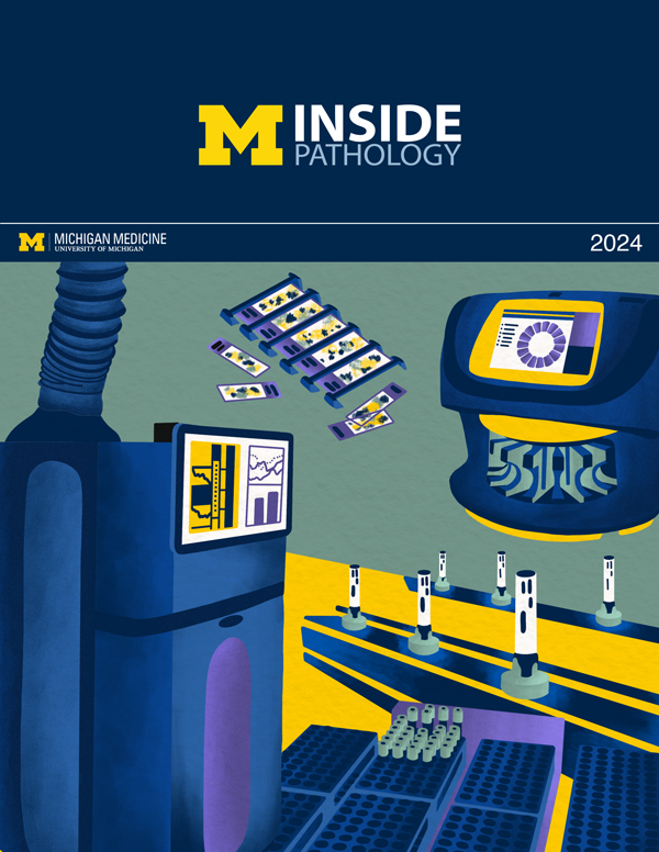
Illustration representing the various machines and processing used within our labs. |
newsletter
INSIDE PATHOLOGYAbout Our NewsletterInside Pathology is an newsletter published by the Chairman's Office to bring news and updates from inside the department's research and to become familiar with those leading it. It is our hope that those who read it will enjoy hearing about those new and familiar, and perhaps help in furthering our research. CONTENTS
|
 ON THE COVER
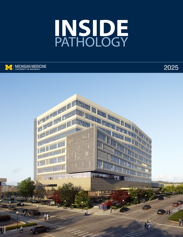
Rendering of the D. Dan and Betty Khn Health Care Pavilion. Credit: HOK |
newsletter
INSIDE PATHOLOGYAbout Our NewsletterInside Pathology is an newsletter published by the Chairman's Office to bring news and updates from inside the department's research and to become familiar with those leading it. It is our hope that those who read it will enjoy hearing about those new and familiar, and perhaps help in furthering our research. CONTENTS
|
MLabs, established in 1985, functions as a portal to provide pathologists, hospitals. and other reference laboratories access to the faculty, staff and laboratories of the University of Michigan Health System’s Department of Pathology. MLabs is a recognized leader for advanced molecular diagnostic testing, helpful consultants and exceptional customer service.