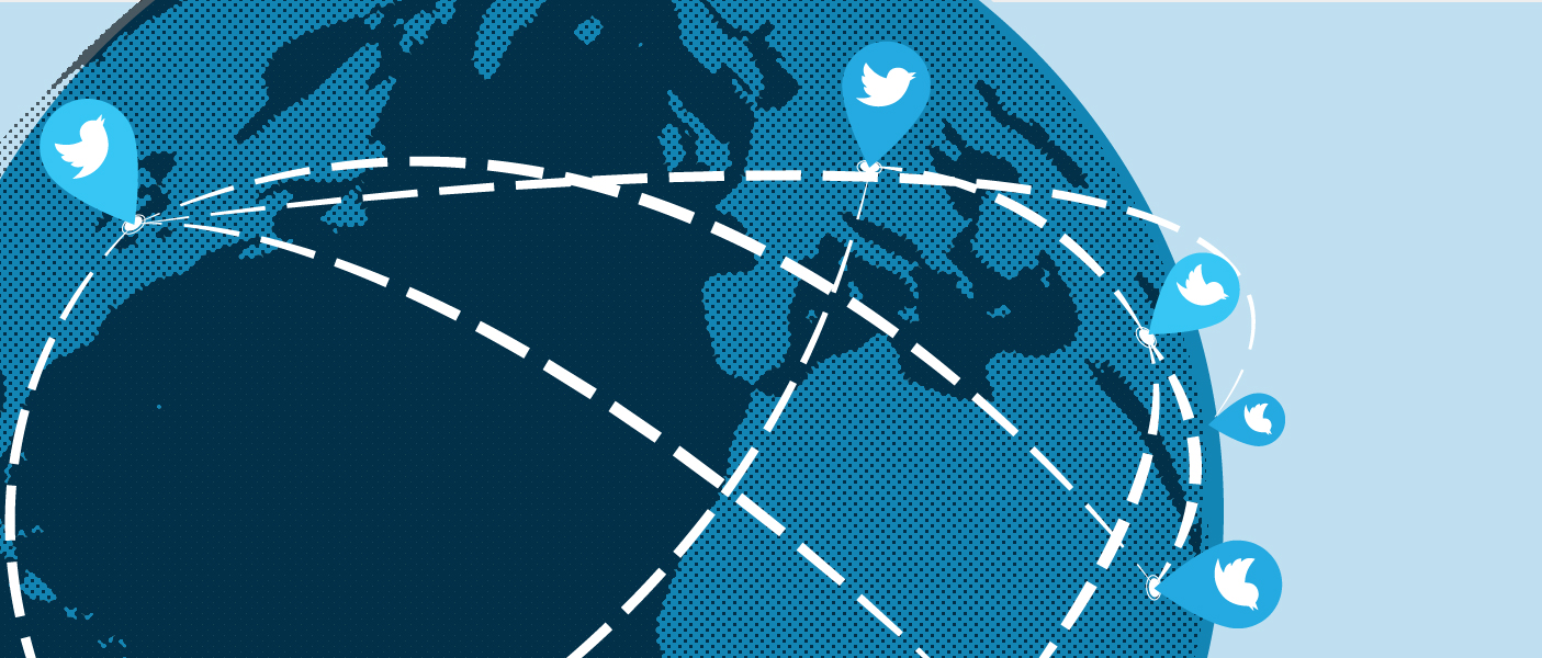
As social media continues to gain popularity among pathologists, users old and new are innovating with new ways to leverage sites like Twitter, Instagram, and Facebook to share new research, discuss interesting cases, and build an encouraging online community that is active 24 hours a day, 7 days a week, 365 days a year. There are infinite possibilities for social media but perhaps the most immediate and significant impact has been in education, including that of pathologists-in-training, and of students considering careers in health care.
The University of Michigan Department of Pathology (@UMichPath) joined Facebook roughly two years ago, and then began Twitter soon after that, and most recently has created profiles on YouTube and Instagram. Across these platforms, @UMichPath has been posting about department events, awards, and news, but it has also begun to correct misconceptions about pathology and reveal the myriad ways in which it is essential to patient care.
The visual nature of pathology lends itself well to image-forward sites like Twitter and Instagram and users constantly share histologic cases for education and discussion. Championed by Drs. Cody Carter and Ellen East, @UMichPath created its own series of cases, dubbed the #DailyDx. The cases from the #DailyDx and others online are popular with trainees looking for more exposure to diagnoses but the cases have found an even wider audience internationally with pathologists in under-resourced countries.
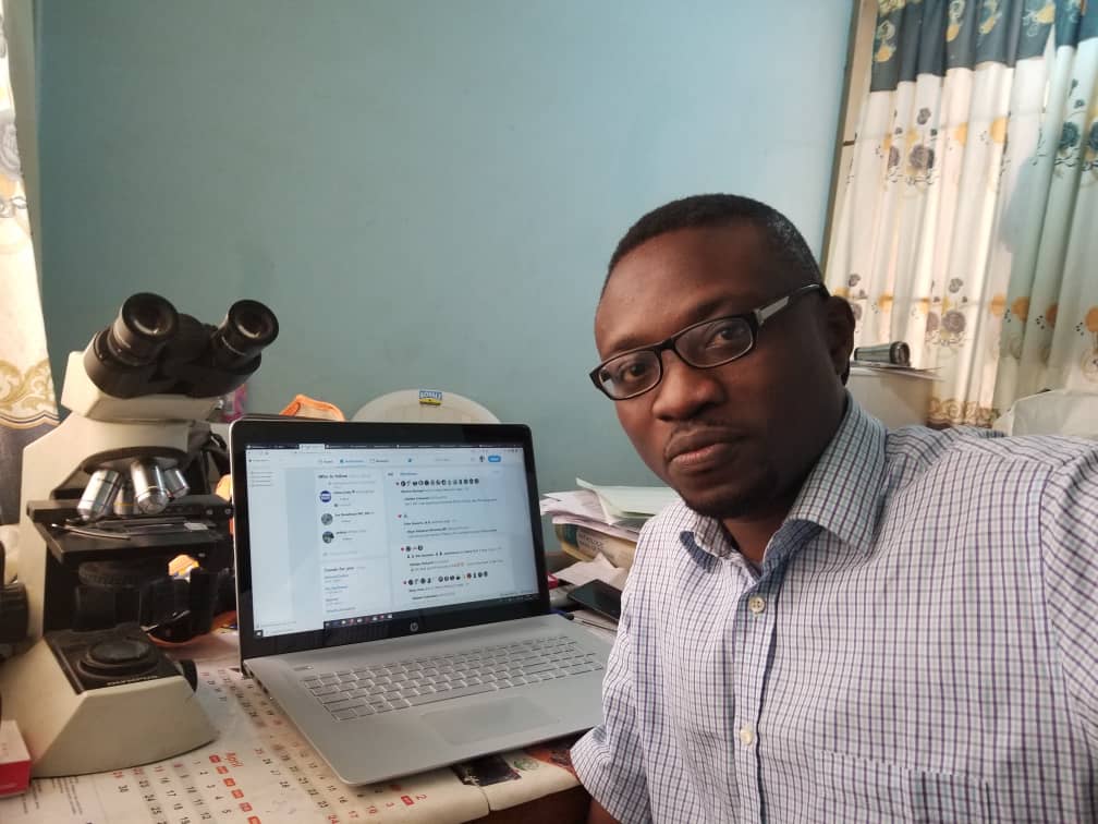
One such pathologist is Dr. Olaleke Folaranmi, who works at the University of Ilorin Teaching Hospital, in Ilorin, Nigeria. Under the handle of @DrGeeONE, Dr. Folaranmi can be found on Twitter every day, offering diagnoses for unknown cases, commenting on new research, and conversing with colleagues about anything pathology-related. He has been active on Twitter for years and was an early fan of the #DailyDx.
“The #DailyDx is a huge source of inspiration to me,” Dr. Folaranmi said. “I never really believed I could get many of these cases until I tried. It serves me as a regular brain teaser and that stimulates me to learn new diseases.” The wide variety of slides shared online by the larger pathology community have been an essential and foundational portion of his learning.
“Without exaggerating,” Dr. Folaranmi stated decisively, “I have learned more pathology from interactions with pathologists and residents on Twitter and Facebook than from physical interactions with people in all my years of training.”
“I have learned more pathology from interactions with pathologists and residents on Twitter and Facebook than from physical interactions with people in all my years of training.”
The hospital where Dr. Folaranmi works is severely underfunded and employs just four pathologists, most of which are bogged down by administrative duties. “We are very limited in what we can offer,” Dr. Folaranmi explained. “It is difficult to get immunohistochemical stains done, and when we do, they are for very few panels.”
This problem is not unique to the University of Ilorin Teaching Hospital. According to Dr. Folaranmi, there are less than 250 pathologists in Nigeria, a country of 190 million people. Although exact numbers are difficult to confirm, a study in 2011 estimated there was 1 anatomic pathologist for every 3 million Nigerians.
With little access to resources or mentors, Dr. Folaranmi turned to social media to enrich his education, even at great personal cost. He took out loans in order to be able to afford a dedicated microscope camera, computer, smartphone with a quality camera, and an expensive data plan. But Dr. Folaranmi will be the first one to tell you it is worth it. “For those of us in underserved countries,” said Dr. Folaranmi, “being on Twitter or other platforms and sharing cases are the only way we can bridge some of the huge knowledge gaps in our training.”
The potential of social media is greater than just advancing the knowledge of those who have already decided on a career in pathology; it also has the ability to inspire the next generation of pathologists. At Hamilton Wenham Regional High School in Hamilton, Massachusetts, digital content shared on social media creates an entirely different educational opportunity.
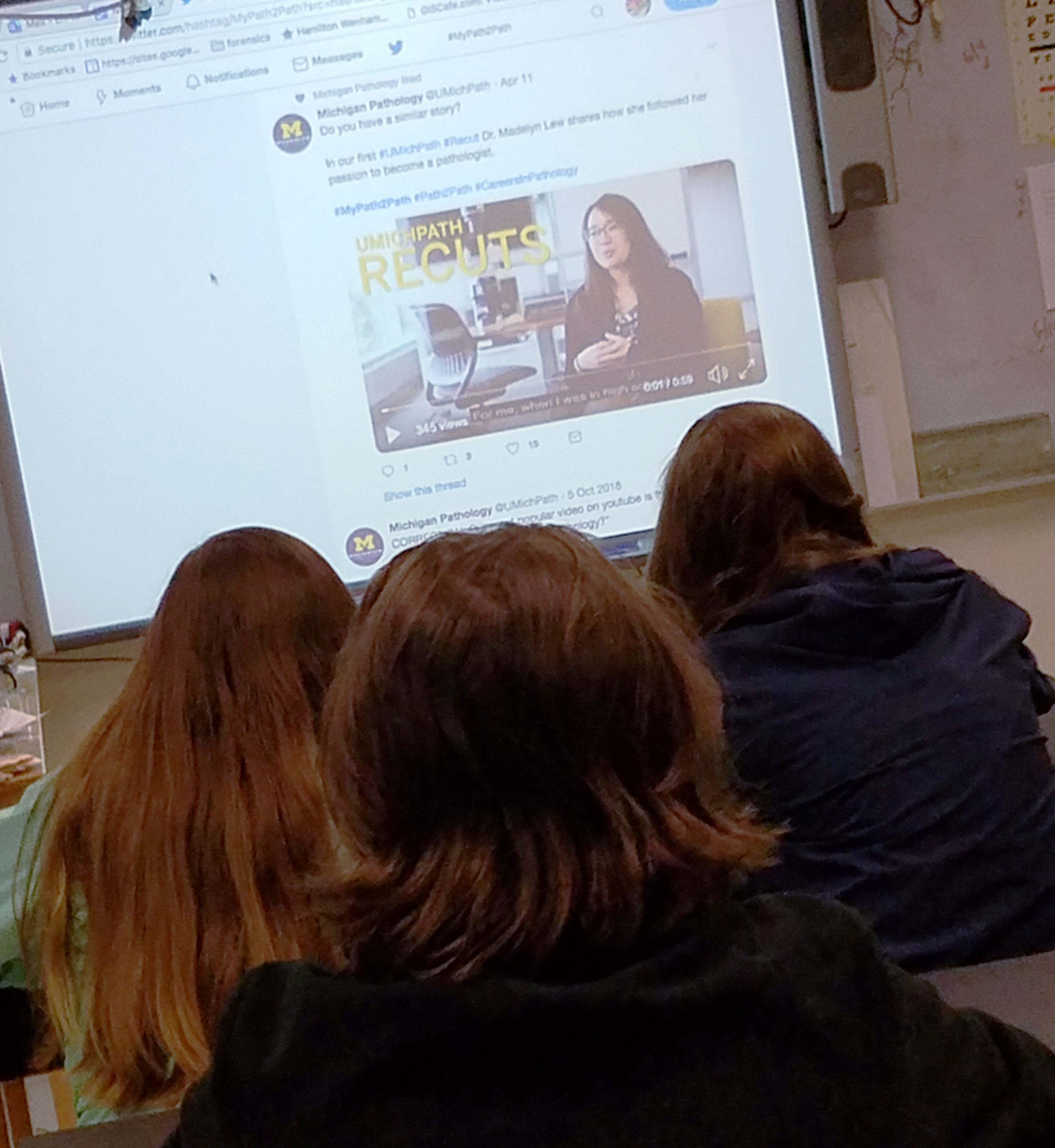
Honors Anatomy and Physiology teacher, Deb Clapp, often begins her class with videos from Twitter and YouTube, including many from @UMichPath. “I love the #MyPath2Path videos.” she explained, “They enable me to bring a real connection to my classroom and help me show all of these different people and the different reasons they decided on pathology as a career.”
Not long ago, Deb was a skeptic of Twitter, but now she sees it as an essential tool for both herself and her students. In the beginning, the school used Twitter as a way for administrators to see what teachers were doing in the classroom and used it as a reference for evaluations. Soon after, parents began following accounts so they could see what their kids were learning in school. But in the past two years, Deb has begun to realize Twitter’s real potential.
 Traditional classroom learning is limited by the knowledge of the teacher and the textbook. Deb used to teach histology using a small number of images of basic cases and a limited knowledge of the pathology of each. Now, through pathology-related social media accounts like @UMichPath, Deb has access to a nearly infinite number of histological images and diagnostic entities and has the ability to reach out to pathologists for help in understanding them.
Instead of simply sharing what they are doing in their classrooms, teachers like Deb are using Twitter as a powerful tool for answering difficult questions. “Students think their teachers know everything,” Deb said, “But I have to be brave enough to say, ‘I don’t know, I’ll look that up for you.’” For answers that are not easily googled, Deb searches Twitter to connect with an expert or to post a question in hopes of crowdsourcing an accurate answer.
“Twitter has made it so much easier for teachers to continue to learn”, Deb explained, “and be able to teach our students with more specific information.” By reaching out to pathologists and other professionals on Twitter, she has made invaluable connections with subject matter experts who are excited to help her enrich her lesson plans. She has even created a “Speaker Series” in which professionals met through Twitter have visited her classroom to talk about their careers.
Deb also simply uses Twitter as a way to find videos that she can use to bookend discussions or assign as homework. She has shown her students the majority of the @UMichPath videos on Twitter and YouTube and because all are available online, students can search the archives themselves. Other teachers also follow Deb on Twitter and can mimic her lessons in their own classrooms.
When students in high school are learning about how fascinating pathology can be, there are no longer misconceptions about the professions that need to be corrected. Deb’s students understand that while some pathologists perform autopsies, most work outside of the morgue diagnosing disease, and some even hold dual appointments in different departments. Even for the students who have no interest in a career in pathology, they now have a general understanding of how cancers and other diseases are diagnosed.
While many people remain skeptical of social media for professional use, those who have embraced it are connecting and finding innovative ways to leverage its ability to disseminate information and level the playing field. It is impossible to know how many pathologists like Dr. Folaranmi are online, learning from this growing network of knowledge, and translating it to better care for their patients. There is no limit to the number of teachers who can show their students @UMichPath videos in the classroom and teach them about careers in pathology. Social media is already changing the landscape of pathology and through educational initiatives and new digital content, we are excited to be at the forefront of this exciting medium.
 ON THE COVER
Breast team reviewing a patient's slide. (From left to right) Ghassan Allo, Fellow; Laura Walters, Clinical Lecturer; Celina Kleer, Professor. See Article |
newsletter
INSIDE PATHOLOGYAbout Our NewsletterInside Pathology is an newsletter published by the Chairman's Office to bring news and updates from inside the department's research and to become familiar with those leading it. It is our hope that those who read it will enjoy hearing about those new and familiar, and perhaps help in furthering our research. CONTENTS
|
 ON THE COVER
Autopsy Technician draws blood while working in the Wayne County morgue. See Article |
newsletter
INSIDE PATHOLOGYAbout Our NewsletterInside Pathology is an newsletter published by the Chairman's Office to bring news and updates from inside the department's research and to become familiar with those leading it. It is our hope that those who read it will enjoy hearing about those new and familiar, and perhaps help in furthering our research. CONTENTS
|
 ON THE COVER
Dr. Sriram Venneti, MD, PhD and Postdoctoral Fellow, Chan Chung, PhD investigate pediatric brain cancer. See Article |
newsletter
INSIDE PATHOLOGYAbout Our NewsletterInside Pathology is an newsletter published by the Chairman's Office to bring news and updates from inside the department's research and to become familiar with those leading it. It is our hope that those who read it will enjoy hearing about those new and familiar, and perhaps help in furthering our research. CONTENTS
|
 ON THE COVER
Director of the Neuropathology Fellowship, Dr. Sandra Camelo-Piragua serves on the Patient and Family Advisory Council. |
newsletter
INSIDE PATHOLOGYAbout Our NewsletterInside Pathology is an newsletter published by the Chairman's Office to bring news and updates from inside the department's research and to become familiar with those leading it. It is our hope that those who read it will enjoy hearing about those new and familiar, and perhaps help in furthering our research. CONTENTS
|
 ON THE COVER
Residents Ashley Bradt (left) and William Perry work at a multi-headed scope in our new facility. |
newsletter
INSIDE PATHOLOGYAbout Our NewsletterInside Pathology is an newsletter published by the Chairman's Office to bring news and updates from inside the department's research and to become familiar with those leading it. It is our hope that those who read it will enjoy hearing about those new and familiar, and perhaps help in furthering our research. CONTENTS
|
 ON THE COVER
Dr. Kristine Konopka (right) instructing residents while using a multi-headed microscope. |
newsletter
INSIDE PATHOLOGYAbout Our NewsletterInside Pathology is an newsletter published by the Chairman's Office to bring news and updates from inside the department's research and to become familiar with those leading it. It is our hope that those who read it will enjoy hearing about those new and familiar, and perhaps help in furthering our research. CONTENTS
|
 ON THE COVER
Patient specimens poised for COVID-19 PCR testing. |
newsletter
INSIDE PATHOLOGYAbout Our NewsletterInside Pathology is an newsletter published by the Chairman's Office to bring news and updates from inside the department's research and to become familiar with those leading it. It is our hope that those who read it will enjoy hearing about those new and familiar, and perhaps help in furthering our research. CONTENTS
|
 ON THE COVER
Dr. Pantanowitz demonstrates using machine learning in analyzing slides. |
newsletter
INSIDE PATHOLOGYAbout Our NewsletterInside Pathology is an newsletter published by the Chairman's Office to bring news and updates from inside the department's research and to become familiar with those leading it. It is our hope that those who read it will enjoy hearing about those new and familiar, and perhaps help in furthering our research. CONTENTS
|
 ON THE COVER
(Left to Right) Drs. Angela Wu, Laura Lamps, and Maria Westerhoff. |
newsletter
INSIDE PATHOLOGYAbout Our NewsletterInside Pathology is an newsletter published by the Chairman's Office to bring news and updates from inside the department's research and to become familiar with those leading it. It is our hope that those who read it will enjoy hearing about those new and familiar, and perhaps help in furthering our research. CONTENTS
|
 ON THE COVER
Illustration representing the various machines and processing used within our labs. |
newsletter
INSIDE PATHOLOGYAbout Our NewsletterInside Pathology is an newsletter published by the Chairman's Office to bring news and updates from inside the department's research and to become familiar with those leading it. It is our hope that those who read it will enjoy hearing about those new and familiar, and perhaps help in furthering our research. CONTENTS
|
 ON THE COVER
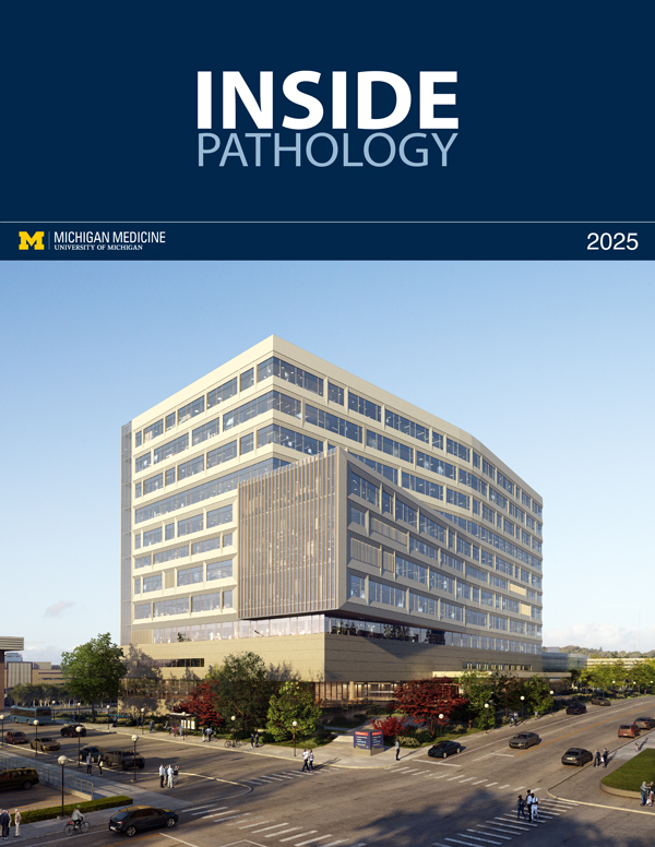
Rendering of the D. Dan and Betty Khn Health Care Pavilion. Credit: HOK |
newsletter
INSIDE PATHOLOGYAbout Our NewsletterInside Pathology is an newsletter published by the Chairman's Office to bring news and updates from inside the department's research and to become familiar with those leading it. It is our hope that those who read it will enjoy hearing about those new and familiar, and perhaps help in furthering our research. CONTENTS
|
MLabs, established in 1985, functions as a portal to provide pathologists, hospitals. and other reference laboratories access to the faculty, staff and laboratories of the University of Michigan Health System’s Department of Pathology. MLabs is a recognized leader for advanced molecular diagnostic testing, helpful consultants and exceptional customer service.