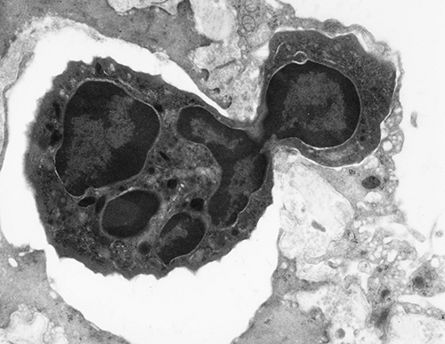
PMN chemotaxingMicroscopes are the workhorses of pathology. What is so different about the electron microscope, and how does it work?
The transmission electron microscope (TEM) was invented by Max Knoll and Ernst Ruska in 1931 in Berlin. Although this first electron microscope succeeded in magnifying an electron beam, it had only a resolving power of 50 nanometers (nm). From this beginning, the field of electron microscopy was born and 50 years later, Ernst Ruska won a Nobel Prize for his efforts.
In 1938 at the University of Toronto, Eli Burton and his students constructed the first electron microscope in the New World. This was higher resolution than Ernst’s TEM and led to what became known as the RCA microscope. Unfortunately, the breakout of World War II set back further developments and it wasn’t until twenty years after the war that commercial TEMs with good resolving power became available.
The electron microscope is similar to the light microscope in that it uses radiation (electron beam vs. light photons) to give a magnified and detailed image of something that the human eye cannot normally see. Electro-magnets are used to focus the electron beam as it passes through the sample in a vacuum. The electron microscope has a much higher spatial resolution than a light microscope (0.2-2.0nm vs. 200nm).
Because the early TEMs used a very high current electron beam that would burn up the samples, they were initially used in metallurgy where the samples could withstand the high voltage. By the 1950s and 60s however, TEM was being utilized in medicine, and especially in pathology. It was discovered that the proteins and lipids in biological samples could be fixed with aldehydes and osmium and the sample could be embedded in plastic. The sample-containing plastic would be thinly sectioned (~90 angstroms), collected on a small 3mm grid, and stained with heavy metals (uranium and lead). The electron beam going through the sample would be stopped by the heavy metals and pass through where there were no heavy metals. These electrons would then be converted to light photons when they struck a phosphorus viewing screen, resulting in an image of the sample. The first cameras added to the TEM used glass plates covered with an electron sensitive coating. Later Kodak made electron image film and today TEMs are completely digital.
Where light microscopy is used to visual groups of cells and tissue patterns, electron microscopy is excellent at giving the pathologist a view of individual cells, and subcellular components. Diagnoses were aided by pathologists being able to see changes in organelles, cytoskeletal and matrix materials, and deposits of subcellular material. The samples currently analyzed by TEM here in the University of Michigan Department of Pathology consist mostly of glomerular diseases of native and transplanted kidneys, muscle and nerve biopsies, skin for CADASIL syndrome, and abdominal fat for systemic amyloidosis. While the TEM was used to diagnose tumors, today that is more routinely done with immunohistochemistry.
Although TEM yields only black and white images, it can take the viewer to places where many people have never gone. Electron microscopes are used in life sciences, nanotechnologies, medicine, biology, forensic analysis, gemology, metallurgy, semiconductor research, quality control, as well as industry and education. Electron microscopes continue to become more powerful and today we can potentially magnify one million times or more. There is a vast world beyond the limits of our vision, and the electron microscope is helping us see it.
Read More About This |
Did You Know? One Cow's Historic Connection to Pathology@umichpath #PathologyDYK |
|
A History of Pathology in 50 Objects Overview of Electron Microscopy |
Intro — Electron Microscopy Electron Microscopy: A Brief History and Review of Current Clinical Application |
2 activated macrophages, lymphocyte and RBC
Basophil
Cilia from lung trachea
Dendritic cell phagocytizing yeast
Dividing lymphocyte
Eosinophil
Glomerulus capillary with fibrin and platelets
Glomerulus capillary with PMNs
Glomerulus (low magnification)
Legionnella bacteria
Lung venial filled with RBC and alveolar macrophage
Lung vessel on grid
Macrophage phagocytizing apoptotic cell
Mitochondria
PMNs phagocytizing Legionnella in lung
Shistosoma egg in lung vessel surrounded by platelets
T4 Phage
Two Lung Capillaries with Macrophage
![]() Breast team reviewing a patient's slide. (From left to right) Ghassan Allo, Fellow; Laura Walters, Clinical Lecturer; Celina Kleer, Professor. See Article 2014Department Chair |
![]()
newsletter
INSIDE PATHOLOGYAbout Our NewsletterInside Pathology is an newsletter published by the Chairman's Office to bring news and updates from inside the department's research and to become familiar with those leading it. It is our hope that those who read it will enjoy hearing about those new and familiar, and perhaps help in furthering our research. CONTENTS
|
![]() Autopsy Technician draws blood while working in the Wayne County morgue. See Article 2016Department Chair |
![]()
newsletter
INSIDE PATHOLOGYAbout Our NewsletterInside Pathology is an newsletter published by the Chairman's Office to bring news and updates from inside the department's research and to become familiar with those leading it. It is our hope that those who read it will enjoy hearing about those new and familiar, and perhaps help in furthering our research. CONTENTS
|
![]() Dr. Sriram Venneti, MD, PhD and Postdoctoral Fellow, Chan Chung, PhD investigate pediatric brain cancer. See Article 2017Department Chair |
![]()
newsletter
INSIDE PATHOLOGYAbout Our NewsletterInside Pathology is an newsletter published by the Chairman's Office to bring news and updates from inside the department's research and to become familiar with those leading it. It is our hope that those who read it will enjoy hearing about those new and familiar, and perhaps help in furthering our research. CONTENTS
|
![]() Director of the Neuropathology Fellowship, Dr. Sandra Camelo-Piragua serves on the Patient and Family Advisory Council. 2018Department Chair |
![]()
newsletter
INSIDE PATHOLOGYAbout Our NewsletterInside Pathology is an newsletter published by the Chairman's Office to bring news and updates from inside the department's research and to become familiar with those leading it. It is our hope that those who read it will enjoy hearing about those new and familiar, and perhaps help in furthering our research. CONTENTS
|
![]() Residents Ashley Bradt (left) and William Perry work at a multi-headed scope in our new facility. 2019Department Chair |
![]()
newsletter
INSIDE PATHOLOGYAbout Our NewsletterInside Pathology is an newsletter published by the Chairman's Office to bring news and updates from inside the department's research and to become familiar with those leading it. It is our hope that those who read it will enjoy hearing about those new and familiar, and perhaps help in furthering our research. CONTENTS
|
![]() Dr. Kristine Konopka (right) instructing residents while using a multi-headed microscope. 2020Department Chair |
![]()
newsletter
INSIDE PATHOLOGYAbout Our NewsletterInside Pathology is an newsletter published by the Chairman's Office to bring news and updates from inside the department's research and to become familiar with those leading it. It is our hope that those who read it will enjoy hearing about those new and familiar, and perhaps help in furthering our research. CONTENTS
|
![]() Patient specimens poised for COVID-19 PCR testing. 2021Department Chair |
![]()
newsletter
INSIDE PATHOLOGYAbout Our NewsletterInside Pathology is an newsletter published by the Chairman's Office to bring news and updates from inside the department's research and to become familiar with those leading it. It is our hope that those who read it will enjoy hearing about those new and familiar, and perhaps help in furthering our research. CONTENTS
|
![]() Dr. Pantanowitz demonstrates using machine learning in analyzing slides. 2022Department Chair |
![]()
newsletter
INSIDE PATHOLOGYAbout Our NewsletterInside Pathology is an newsletter published by the Chairman's Office to bring news and updates from inside the department's research and to become familiar with those leading it. It is our hope that those who read it will enjoy hearing about those new and familiar, and perhaps help in furthering our research. CONTENTS
|
![]() (Left to Right) Drs. Angela Wu, Laura Lamps, and Maria Westerhoff. 2023Department Chair |
![]()
newsletter
INSIDE PATHOLOGYAbout Our NewsletterInside Pathology is an newsletter published by the Chairman's Office to bring news and updates from inside the department's research and to become familiar with those leading it. It is our hope that those who read it will enjoy hearing about those new and familiar, and perhaps help in furthering our research. CONTENTS
|
![]() Illustration representing the various machines and processing used within our labs. 2024Department Chair |
![]()
newsletter
INSIDE PATHOLOGYAbout Our NewsletterInside Pathology is an newsletter published by the Chairman's Office to bring news and updates from inside the department's research and to become familiar with those leading it. It is our hope that those who read it will enjoy hearing about those new and familiar, and perhaps help in furthering our research. CONTENTS
|
MLabs, established in 1985, functions as a portal to provide pathologists, hospitals. and other reference laboratories access to the faculty, staff and laboratories of the University of Michigan Health System’s Department of Pathology. MLabs is a recognized leader for advanced molecular diagnostic testing, helpful consultants and exceptional customer service.