A new approach to the practice of surgical pathology for brain tumor patients could make for a powerful combination: more accurate, safer and more efficient operations.
Neurosurgeons and pathologists at Michigan Medicine are the first to execute stimulated Raman histology, a method that improves speed and diagnostic efficiency, in an operating room. They detail the advance in a new Nature Biomedical Engineering paper.
The researchers imaged tissue from 101 neurosurgical patients using conventional methods and the new method. Both techniques, they found, produced accurate results but the new method was much faster.
That, if applied widely, could change the pace and structure of an operation.
“By achieving excellent image quality in fresh tissues, we're able to make a diagnosis during surgery,” says first author Daniel A. Orringer, M.D., assistant professor of neurosurgery at the University of Michigan Medical School. “This eliminates the lengthy process of sending tissues out of the OR for processing and interpretation.”
Today’s workflow for determining a diagnosis during an operation requires the surgeon wait for 30 to 40 minutes while tissue is sent to a dedicated pathology lab for processing, sectioning, staining, mounting and interpretation. The entire team in the operating room may be idle while waiting for pathology results, Orringer says.
A more efficient surgical procedure would save money by requiring less time in the operating room.
“Our technique may disrupt the intraoperative diagnosis process in a great way, reducing it from a 30-minute process to about 3 minutes,” Orringer says. “Initially, we developed this technology as a means of helping surgeons detect microscopic tumor, but we found the technology was capable of much more than guiding surgery.”
Stimulated Raman scattering microscopy, the technology behind SRH, was developed in 2008, but the hazardous lasers it involved made it unsuitable for use in an operating room. A clinical version has now been developed and tested in the operating room for more than a year at U-M, with the fiber-laser-based microscope mounted right onto a clinical cart that plugs into the wall.
To interpret the samples, researchers developed SRH, which creates images familiar to those currently in use.
SRH uses virtual coloring to highlight the cellular and architectural features of brain tumors, with a result resembling traditional staining. The pathologist is then able to differentiate the tumor tissue from normal brain as usual.
“It’s very similar to what we currently do in our intraoperative diagnosis, with the exception that the tissue is fresh, has not been processed or stained,” says senior author Sandra Camelo-Piragua, M.D., assistant professor of pathology at the U-M Medical School.
In the Nature Biomedical Engineering study, neuropathologists were given 30 specimen samples, processed via SRH or traditional methods. They were told the same information about each patient’s medical history and the location of the tumor and asked to make a diagnosis.
Those pathologists, the U-M researchers found, were equally likely to make a correct diagnosis whether they used SRH or conventional slides.
“SRH imaging will ensure that appropriate and good quality tissue is collected to reach our ultimate goal: accurate diagnosis,” Camelo-Piragua says.
As Orringer and his team continue to improve this imaging technology, they’re also teaching a computer how to use SRH images to make diagnoses.
They built and validated a machine learning process that was able to predict brain tumor subtype with 90 percent accuracy in a subset of 30 patient samples.
“The more we feed the computer, the more accurate its diagnoses will become,” Orringer says.
Using SRH might also improve the workflow for facilities without access to expert neuropathologists. Orringer notes that smaller hospitals may be able to partner with larger systems that do have access, since there are fewer than 800 board-certified neuropathologists compared to the approximately 1,400 U.S. institutions performing brain surgery.
“Bringing the SRH to smaller hospitals would extend their capabilities because the images can be interpreted remotely,” he says. Sample preparation is minimal and the SRH could quickly deliver virtual histologic sections to aid diagnosis remotely.
The next step is a large-scale clinical trial, with an eventual goal of showing equivalence between SRH technique for making diagnoses, Orringer says. The prototype system is currently intended for research use only.
—
Additional authors from Michigan Medicine include Balaji Pandian, Yashar S. Niknafs, Todd C. Hollon, Julianne Boyle, Spencer Lewis, Mia Garrard, Shawn L. Hervey-Jumper, Hugh J. L. Garton, Cormac O. Maher, Jason A. Heth, Oren Sagher, D. Andrew Wilkinson, Sriram Venneti, Kathryn A. McFadden, Amanda Fisher-Hubbard, Andrew P. Lieberman and Timothy D. Johnson. Matija Snuderl is from New York University. Shakti H. Ramkissoon is from Harvard Medical School and the Dana Farber Cancer Institute. X. Sunney Xie is from Harvard University. Jay K. Trautman and Christian W. Freudiger are from Invenio Imaging.
Disclosure: Orringer and co-author Xie are advisers and shareholders of Invenio Imaging Inc., a company developing SRS microscopy systems. Co-author Freudiger is an employee and shareholder of Invenio Imaging, Inc.
The University of Michigan, Harvard University and Invenio Imaging have intellectual property used in this research.
 ON THE COVER
Breast team reviewing a patient's slide. (From left to right) Ghassan Allo, Fellow; Laura Walters, Clinical Lecturer; Celina Kleer, Professor. See Article |
newsletter
INSIDE PATHOLOGYAbout Our NewsletterInside Pathology is an newsletter published by the Chairman's Office to bring news and updates from inside the department's research and to become familiar with those leading it. It is our hope that those who read it will enjoy hearing about those new and familiar, and perhaps help in furthering our research. CONTENTS
|
 ON THE COVER
Autopsy Technician draws blood while working in the Wayne County morgue. See Article |
newsletter
INSIDE PATHOLOGYAbout Our NewsletterInside Pathology is an newsletter published by the Chairman's Office to bring news and updates from inside the department's research and to become familiar with those leading it. It is our hope that those who read it will enjoy hearing about those new and familiar, and perhaps help in furthering our research. CONTENTS
|
 ON THE COVER
Dr. Sriram Venneti, MD, PhD and Postdoctoral Fellow, Chan Chung, PhD investigate pediatric brain cancer. See Article |
newsletter
INSIDE PATHOLOGYAbout Our NewsletterInside Pathology is an newsletter published by the Chairman's Office to bring news and updates from inside the department's research and to become familiar with those leading it. It is our hope that those who read it will enjoy hearing about those new and familiar, and perhaps help in furthering our research. CONTENTS
|
 ON THE COVER
Director of the Neuropathology Fellowship, Dr. Sandra Camelo-Piragua serves on the Patient and Family Advisory Council. |
newsletter
INSIDE PATHOLOGYAbout Our NewsletterInside Pathology is an newsletter published by the Chairman's Office to bring news and updates from inside the department's research and to become familiar with those leading it. It is our hope that those who read it will enjoy hearing about those new and familiar, and perhaps help in furthering our research. CONTENTS
|
 ON THE COVER
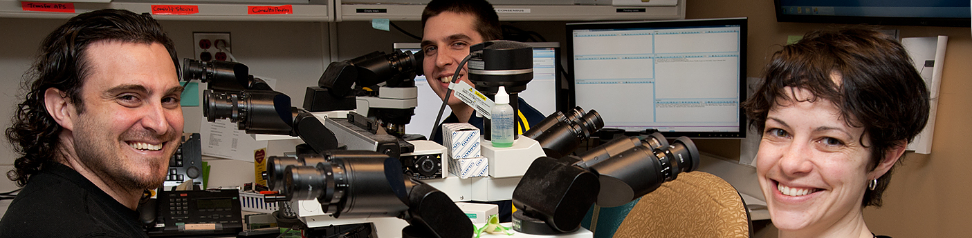
Residents Ashley Bradt (left) and William Perry work at a multi-headed scope in our new facility. |
newsletter
INSIDE PATHOLOGYAbout Our NewsletterInside Pathology is an newsletter published by the Chairman's Office to bring news and updates from inside the department's research and to become familiar with those leading it. It is our hope that those who read it will enjoy hearing about those new and familiar, and perhaps help in furthering our research. CONTENTS
|
 ON THE COVER
Dr. Kristine Konopka (right) instructing residents while using a multi-headed microscope. |
newsletter
INSIDE PATHOLOGYAbout Our NewsletterInside Pathology is an newsletter published by the Chairman's Office to bring news and updates from inside the department's research and to become familiar with those leading it. It is our hope that those who read it will enjoy hearing about those new and familiar, and perhaps help in furthering our research. CONTENTS
|
 ON THE COVER
Patient specimens poised for COVID-19 PCR testing. |
newsletter
INSIDE PATHOLOGYAbout Our NewsletterInside Pathology is an newsletter published by the Chairman's Office to bring news and updates from inside the department's research and to become familiar with those leading it. It is our hope that those who read it will enjoy hearing about those new and familiar, and perhaps help in furthering our research. CONTENTS
|
 ON THE COVER
Dr. Pantanowitz demonstrates using machine learning in analyzing slides. |
newsletter
INSIDE PATHOLOGYAbout Our NewsletterInside Pathology is an newsletter published by the Chairman's Office to bring news and updates from inside the department's research and to become familiar with those leading it. It is our hope that those who read it will enjoy hearing about those new and familiar, and perhaps help in furthering our research. CONTENTS
|
 ON THE COVER
(Left to Right) Drs. Angela Wu, Laura Lamps, and Maria Westerhoff. |
newsletter
INSIDE PATHOLOGYAbout Our NewsletterInside Pathology is an newsletter published by the Chairman's Office to bring news and updates from inside the department's research and to become familiar with those leading it. It is our hope that those who read it will enjoy hearing about those new and familiar, and perhaps help in furthering our research. CONTENTS
|
 ON THE COVER
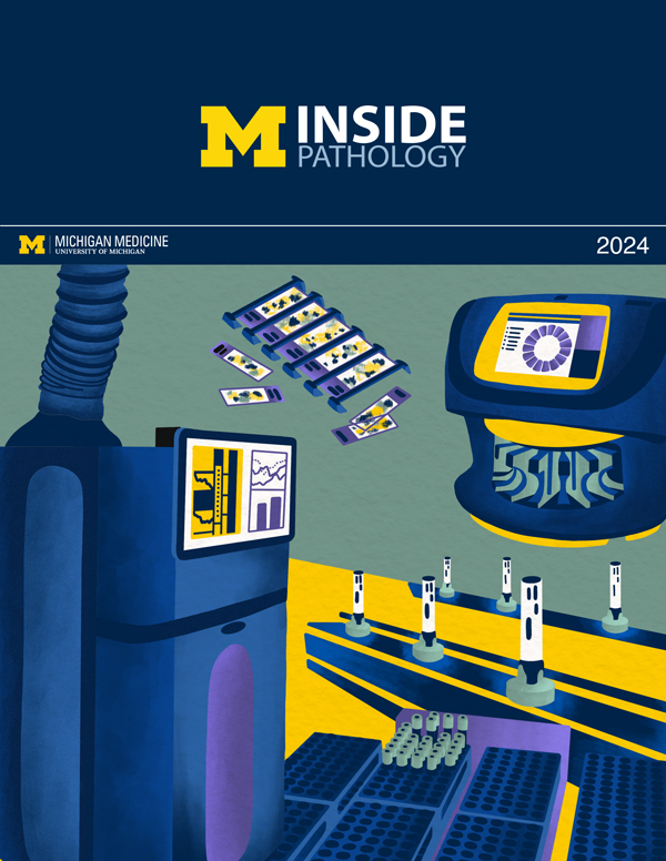
Illustration representing the various machines and processing used within our labs. |
newsletter
INSIDE PATHOLOGYAbout Our NewsletterInside Pathology is an newsletter published by the Chairman's Office to bring news and updates from inside the department's research and to become familiar with those leading it. It is our hope that those who read it will enjoy hearing about those new and familiar, and perhaps help in furthering our research. CONTENTS
|
MLabs, established in 1985, functions as a portal to provide pathologists, hospitals. and other reference laboratories access to the faculty, staff and laboratories of the University of Michigan Health System’s Department of Pathology. MLabs is a recognized leader for advanced molecular diagnostic testing, helpful consultants and exceptional customer service.