Using Artificial Intelligence to predict ERG Gene Fusion in Prostate Cancer
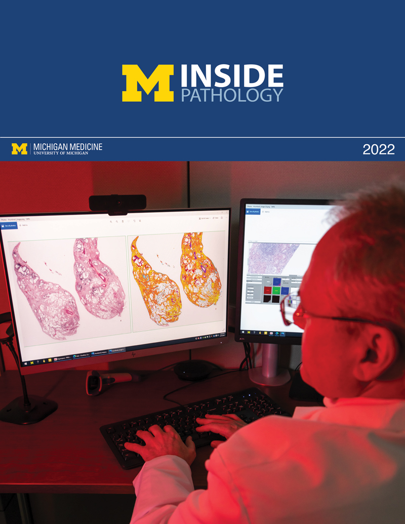
By Lynn McCain | May 10 2022The role of artificial intelligence (AI) in healthcare continues to expand. In a recent issue of BMC Cancer, Dr. Vipulkumar Dadhania (first author) and colleagues published a result of their study Leveraging artificial intelligence to predict ERG gene fusion status in prostate cancer. The expert team from the Michigan Center for Translational Pathology developed a deep-learning-based model to predict ERG genomic rearrangements in prostatic adenocarcinomas using only H&E-stained digital slides. Their AI models were accurate at 78.6%-79.7%, depending on the magnification, with a 20x magnification the most accurate. Sensitivity was found to be at 75% across magnification levels (10x, 20x, 40x) and specificity ranged from 81.7% (10x and 40x) to 83.1% (20x).
Senior Author Rohit Mehra, MD explained, “We plan to look at larger datasets and also to collaborate across institutions to find out how this model holds strength in follow up studies. We will also interrogate which image features specifically enable this H&E-based detection of ERG genomic rearrangements in prostate cancer, and will develop deeper and more refined algorithms to improve sensitivity and specificity of this detection.”
Currently, pathologists must do follow-up testing when they suspect an ERG genomic rearrangement may be present, including next generation sequencing (NGS) genomic studies, immunohistochemical tests (IHC), and fluorescent in situ hybridization (FISH) studies, which take significant amounts of time and require larger tissue samples. “We now have tools to do things we couldn’t do before with a microscope,” commented Liron Pantanowitz, Director of Anatomic Pathology. “This opens up a whole new world for pathologists to make better predictions, prognosis and discovery.”
Mehra continued, “This AI based testing opens up avenues to look at genomic rearrangements using elemental histologic techniques (like H&E staining), and carries promise to save time, tissue, effort, and resources for ancillary (genomic) diagnostic testing for patients. In summary our results suggest that automatically derived image features can capture subtle morphological differences between ERG-rearrarranged and ERG-negative prostate cancer cases.”
 ON THE COVER
 ON THE COVER
 ON THE COVER
 ON THE COVER
 ON THE COVER
 ON THE COVER
 ON THE COVER
 ON THE COVER
 ON THE COVER
 ON THE COVER
 ON THE COVER
