
For patient advocate Michele Mitchell, breast cancer was the invisible enemy she never saw coming. Her journey began with a diagnosis of Stage 1A invasive ductal carcinoma.
At the University of Michigan Comprehensive Cancer Center, she underwent a lumpectomy, chemotherapy, radiation, an additional surgery, and several years of anti-hormonal therapy. Now in remission, Michele shares her story to inspire others to advocate for themselves and to honor the many professionals who played a role in her care, especially the pathologists and laboratory experts whose precise diagnostic work made her treatment possible.
When Michele was first diagnosed, she never saw her pathology report or the slides that revealed her disease. She was told that her breast cancer had been caught early. What she knew was that it was invasive ductal carcinoma, stage 1, small, with no lymph node involvement. Her oncologist reassured her that her prognosis looked “good.” Over the years, she conducted her own research and learned a great deal, yet many questions remained. She wanted to see the cancer for herself to understand what had once been an unseen threat.
A decade after her diagnosis, Michele reached out to Dr. Jeffrey Myers, a pathologist at the University of Michigan, who graciously fulfilled that wish.
“I never thought I could see my cancer,” she recalled. “I really wanted to see the enemy.”
Dr. Myers began by showing her slides of normal breast tissue before displaying a digital image of her own tumor. He explained how the slides were stained and how those colors revealed the tumor’s unique characteristics. Michele’s cancer was classified as luminal A estrogen and progesterone receptor positive, HER2 negative. He also introduced her to the concept of tumor grading, something she had never heard of before.
Dr. Myers explained that the black dots she saw on the screen were cancer cells, and that the characteristics of individual cells helped determine the tumor’s grade, an indicator of its aggressiveness. Michele learned that her tumor was grade II, meaning it grew faster than low-grade cancers but was not the most aggressive type. “When he showed me the black dots on the screen, my cancer, I started to cry,” she said. “He came over and hugged me.”
The encounter was emotional and enlightening. For the first time, she could see the reality of what had once only been a diagnosis on paper. “The sheer size of the enemy,” she stated, “is what stays with me from that experience.”
Even after many years, she sometimes wonders whether some of those cells still linger somewhere inside her. Yet, what she remembers most is the compassion behind that moment. “In a world filled with barcodes, sterile instruments, and starched white lab coats, Dr. Myers extended a warm touch.”
The experience transformed Michele, both in her perception of her health and her future. Determined to take control, she made significant lifestyle changes: eliminating red meat and soy, adopting a consistent exercise routine, and losing over 100 pounds. “I’m convinced these changes will have a positive effect on my health and my cancer journey long term,” she reflected.
Today, Michele keeps a framed picture of her cancer on her dresser, a daily reminder of resilience, vigilance, and hope. “Seeing it reminded me that even something so small can change your life,” she said. “But it also showed me how much power I have to fight back.”
Flashforward to 2025, Michele's year has been challenging after a frightening biopsy scare and a diagnosis of hypercalcemia and osteoporosis. Her endocrinologist ordered blood work and urine studies, both of which passed through the department of pathology labs. Over a four-month period, three urine studies revealed high levels of phosphorus, uric acid, magnesium, and calcium originating from her kidneys.
To seek clarity and understanding of her test results, Michele contacted Dr. David Keren, a professor of clinical pathology. “Dr. Keren spent a significant amount of time with me, thoroughly explaining my studies. He also took the time to explain the NTX telapeptide test, which had been sent to an external reference lab for analysis,” Michele said. She also noted that Dr. Keren spent more time with her than her endocrinologist.
With the information provided by Dr. Keren, the next step was to understand hypercalcemia and early onset osteoporosis. Through research, Michele discovered that a drug she was taking for migraine management, Topomax, has a direct link to both conditions. Additionally, she unearthed that this medication was also linked to her severe leg cramps.
“This breakthrough would not have been possible without Dr. Keren’s compassionate and easily comprehensible explanation of my urine studies,” she said. “Without him, I would have never made this connection.”
“I will forever be grateful to Drs. Myers and Keren. They have truly made a difference in my life.”
 However, this is not where the story ends. Bill, Michele’s husband, is a two-time melanoma survivor. Bill came to Michigan Medicine and received two “picture frame” procedures at the Rogel Cancer Center.
This procedure removed a portion of Bill’s skin around his cancer for a pathologist to review. Dr. Douglas Fullen, now retired from U-M, was the pathologist assigned to the case. “Dr. Fullen promptly informed us that the initial procedure did not achieve clear margins,” she stated. “Upon further examination, he discovered melanoma which led to the second procedure.”
With the second specimen gathered, Dr. Fullen’s analysis and results were turned around and delivered to Bill and Michele. This provided them with the information needed to meet with an Ear, Nose, and Throat specialist to remove the cancer and reconstruct Bill’s cheek effectively.
“We thank Dr. Fullen for his actions and compassionate delivery of diagnosis to Bill and me, while we navigated our health challenges,” Mitchell said. “While we are still navigating our health challenges, it brings us comfort to know that these exceptional pathologists are on our side.”
Throughout the years of navigating challenging health scares and topics, Michele now dedicates her time to being a patient advocate. She enthusiastically shares her positive patient experiences at Michigan Medicine and with the Department of Pathology broadly, noting that Michigan Medicine is the only healthcare institution she trusts for her family's care.
 Through her advocacy work, Michele has collaborated with the American Society for Clinical Pathology, the Digital Pathology Association, the Michigan Cancer Consortium, and other organizations. In addition to her work with these groups, Mitchell shares her story at numerous conferences across the country and paints in an effort to help advocate for patients and solidify her story.
Her artwork, “Renewal” and “The Patient Behind the Lens”, have been featured in The Pathologist and Lilly Oncology on Canvas. “Renewal” was even selected for the Lilly Oncology on Canvas Hope Murals Project and traveled across the US and Puerto Rico before returning home to Michigan.
“I hold no formal power, but I have a voice. And I will keep using it until every patient sees, and every pathologist remembers, the life behind the lens.”
Here at Michigan Medicine, we do just that: remember every life behind the lens.
This International Pathology Day, consider supporting the vital work being done in our Department. Learn more about how your support helps here.
 ON THE COVER
Breast team reviewing a patient's slide. (From left to right) Ghassan Allo, Fellow; Laura Walters, Clinical Lecturer; Celina Kleer, Professor. See Article |
newsletter
INSIDE PATHOLOGYAbout Our NewsletterInside Pathology is an newsletter published by the Chairman's Office to bring news and updates from inside the department's research and to become familiar with those leading it. It is our hope that those who read it will enjoy hearing about those new and familiar, and perhaps help in furthering our research. CONTENTS
|
 ON THE COVER
Autopsy Technician draws blood while working in the Wayne County morgue. See Article |
newsletter
INSIDE PATHOLOGYAbout Our NewsletterInside Pathology is an newsletter published by the Chairman's Office to bring news and updates from inside the department's research and to become familiar with those leading it. It is our hope that those who read it will enjoy hearing about those new and familiar, and perhaps help in furthering our research. CONTENTS
|
 ON THE COVER
Dr. Sriram Venneti, MD, PhD and Postdoctoral Fellow, Chan Chung, PhD investigate pediatric brain cancer. See Article |
newsletter
INSIDE PATHOLOGYAbout Our NewsletterInside Pathology is an newsletter published by the Chairman's Office to bring news and updates from inside the department's research and to become familiar with those leading it. It is our hope that those who read it will enjoy hearing about those new and familiar, and perhaps help in furthering our research. CONTENTS
|
 ON THE COVER
Director of the Neuropathology Fellowship, Dr. Sandra Camelo-Piragua serves on the Patient and Family Advisory Council. |
newsletter
INSIDE PATHOLOGYAbout Our NewsletterInside Pathology is an newsletter published by the Chairman's Office to bring news and updates from inside the department's research and to become familiar with those leading it. It is our hope that those who read it will enjoy hearing about those new and familiar, and perhaps help in furthering our research. CONTENTS
|
 ON THE COVER
Residents Ashley Bradt (left) and William Perry work at a multi-headed scope in our new facility. |
newsletter
INSIDE PATHOLOGYAbout Our NewsletterInside Pathology is an newsletter published by the Chairman's Office to bring news and updates from inside the department's research and to become familiar with those leading it. It is our hope that those who read it will enjoy hearing about those new and familiar, and perhaps help in furthering our research. CONTENTS
|
 ON THE COVER
Dr. Kristine Konopka (right) instructing residents while using a multi-headed microscope. |
newsletter
INSIDE PATHOLOGYAbout Our NewsletterInside Pathology is an newsletter published by the Chairman's Office to bring news and updates from inside the department's research and to become familiar with those leading it. It is our hope that those who read it will enjoy hearing about those new and familiar, and perhaps help in furthering our research. CONTENTS
|
 ON THE COVER
Patient specimens poised for COVID-19 PCR testing. |
newsletter
INSIDE PATHOLOGYAbout Our NewsletterInside Pathology is an newsletter published by the Chairman's Office to bring news and updates from inside the department's research and to become familiar with those leading it. It is our hope that those who read it will enjoy hearing about those new and familiar, and perhaps help in furthering our research. CONTENTS
|
 ON THE COVER
Dr. Pantanowitz demonstrates using machine learning in analyzing slides. |
newsletter
INSIDE PATHOLOGYAbout Our NewsletterInside Pathology is an newsletter published by the Chairman's Office to bring news and updates from inside the department's research and to become familiar with those leading it. It is our hope that those who read it will enjoy hearing about those new and familiar, and perhaps help in furthering our research. CONTENTS
|
 ON THE COVER
(Left to Right) Drs. Angela Wu, Laura Lamps, and Maria Westerhoff. |
newsletter
INSIDE PATHOLOGYAbout Our NewsletterInside Pathology is an newsletter published by the Chairman's Office to bring news and updates from inside the department's research and to become familiar with those leading it. It is our hope that those who read it will enjoy hearing about those new and familiar, and perhaps help in furthering our research. CONTENTS
|
 ON THE COVER
Illustration representing the various machines and processing used within our labs. |
newsletter
INSIDE PATHOLOGYAbout Our NewsletterInside Pathology is an newsletter published by the Chairman's Office to bring news and updates from inside the department's research and to become familiar with those leading it. It is our hope that those who read it will enjoy hearing about those new and familiar, and perhaps help in furthering our research. CONTENTS
|
 ON THE COVER
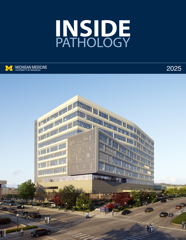
Rendering of the D. Dan and Betty Khn Health Care Pavilion. Credit: HOK |
newsletter
INSIDE PATHOLOGYAbout Our NewsletterInside Pathology is an newsletter published by the Chairman's Office to bring news and updates from inside the department's research and to become familiar with those leading it. It is our hope that those who read it will enjoy hearing about those new and familiar, and perhaps help in furthering our research. CONTENTS
|
MLabs, established in 1985, functions as a portal to provide pathologists, hospitals. and other reference laboratories access to the faculty, staff and laboratories of the University of Michigan Health System’s Department of Pathology. MLabs is a recognized leader for advanced molecular diagnostic testing, helpful consultants and exceptional customer service.