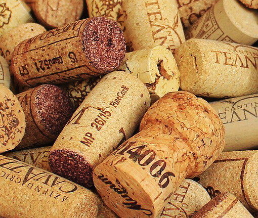
Many of us likely remember studying the cell structure from a diagram in our school's science class; learning about the nucleus, the nuclear membrane, and cell membrane and how it contributes to the basic structure and function of all living organisms. But did you know that the term "cell" came from the material used to plug your wine bottle?
After the first compound microscope was created in the 1590's, its use was more of a novelty with a maximum magnification of 9x. It wasn't until the mid-1600's that the microscope gained interest in the scientific community with magnification reaching 50x. Because of a commission by King Charles II of England, 26 year old English scientist, Robert Hooke, took on an assignment to create a series of microscopical studies.
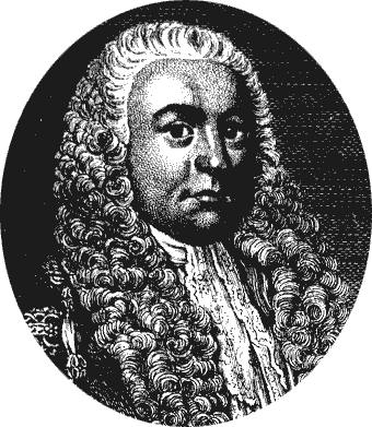
Portrait of Robert Hooke.Hooke later published Micrographia: or Some Physiological Descriptions of Miniature Bodies Made by Magnifying Glasses in 1665 that outlined his observations of material and living things he made with a primitive microscope. It proved to be the first of many things: the first major publication of the Royal Society, and the first scientific best-seller that inspired a wide public interest in the new science of microscopy. Among his observations were insects, fabric, and fossils that he illustrated himself; often illustrating them within a circle, reflecting his viewpoint from the microscope's lens. His curiosity even took him to observing a louse, a wingless parasitic insect, feeding on his hand to observe how his blood traveled into the louse.
But he also observed plant material which included bark from the tree Quercus suber. This bark, also known as "cork", is light and soft which winemakers use to stop their wine-bottles. Interestingly, while observing a sample of cork under his microscope, he used the word 'cell', Latin meaning "small room", to describe the empty spaces contained by walls within the dead tissue, because it reminded him of the small, simple rooms inside Catholic and Orthodox monasteries and Buddhist vihara of that time.
Though he did not fully understand the meaning of his discovery, the book's publication inspired scientists, no doubt with its beautiful illustrations and curious details, to explore microscopy and led to the now universally accepted cell theory. The first living cells were not seen until 1670 by Anton van Leeuwenhoek, a Dutch biologist, and in 1833, Robert Brown discovered the nucleus, the center, of cells in plants. But because of Robert Hooke's accurate description, the term "cell" stuck.
Read More About This |
Did You Know? Logwood - A History of Palettes, Pirates, and Pathology
@umichpath |
|
A History of Pathology in 50 Objects
Robert Hooke and The Discovery of the Cell
History: Robert Hooke |
History of the Microscope
A Brief History of Microbiology |
Cork - Schem. XI - Robert Hooke Micrographia
Illustration of the Microscope Robert Hooke modified and used during his research for Micrographia
Flea - Robert Hooke Micrographia
Ant - Schem. XXXII - Robert Hooke Micrographia
Closeup of a Fly’s Head - Schem. XXIV - Robert Hooke Micrographia
Gnat - Schem. XXIX - Robert Hooke Micrographia
Louse - Schem. XXXV - Robert Hooke Micrographia
Kettering-Stone - Schem. IX - Robert Hooke Micrographia
Modern scanning of Pollen from an electron microscope. Provided by Dartmouth College Electron Microscope Facility.
![]() Breast team reviewing a patient's slide. (From left to right) Ghassan Allo, Fellow; Laura Walters, Clinical Lecturer; Celina Kleer, Professor. See Article 2014Department Chair |
![]()
newsletter
INSIDE PATHOLOGYAbout Our NewsletterInside Pathology is an newsletter published by the Chairman's Office to bring news and updates from inside the department's research and to become familiar with those leading it. It is our hope that those who read it will enjoy hearing about those new and familiar, and perhaps help in furthering our research. CONTENTS
|
![]() Autopsy Technician draws blood while working in the Wayne County morgue. See Article 2016Department Chair |
![]()
newsletter
INSIDE PATHOLOGYAbout Our NewsletterInside Pathology is an newsletter published by the Chairman's Office to bring news and updates from inside the department's research and to become familiar with those leading it. It is our hope that those who read it will enjoy hearing about those new and familiar, and perhaps help in furthering our research. CONTENTS
|
![]() Dr. Sriram Venneti, MD, PhD and Postdoctoral Fellow, Chan Chung, PhD investigate pediatric brain cancer. See Article 2017Department Chair |
![]()
newsletter
INSIDE PATHOLOGYAbout Our NewsletterInside Pathology is an newsletter published by the Chairman's Office to bring news and updates from inside the department's research and to become familiar with those leading it. It is our hope that those who read it will enjoy hearing about those new and familiar, and perhaps help in furthering our research. CONTENTS
|
![]() Director of the Neuropathology Fellowship, Dr. Sandra Camelo-Piragua serves on the Patient and Family Advisory Council. 2018Department Chair |
![]()
newsletter
INSIDE PATHOLOGYAbout Our NewsletterInside Pathology is an newsletter published by the Chairman's Office to bring news and updates from inside the department's research and to become familiar with those leading it. It is our hope that those who read it will enjoy hearing about those new and familiar, and perhaps help in furthering our research. CONTENTS
|
![]() Residents Ashley Bradt (left) and William Perry work at a multi-headed scope in our new facility. 2019Department Chair |
![]()
newsletter
INSIDE PATHOLOGYAbout Our NewsletterInside Pathology is an newsletter published by the Chairman's Office to bring news and updates from inside the department's research and to become familiar with those leading it. It is our hope that those who read it will enjoy hearing about those new and familiar, and perhaps help in furthering our research. CONTENTS
|
![]() Dr. Kristine Konopka (right) instructing residents while using a multi-headed microscope. 2020Department Chair |
![]()
newsletter
INSIDE PATHOLOGYAbout Our NewsletterInside Pathology is an newsletter published by the Chairman's Office to bring news and updates from inside the department's research and to become familiar with those leading it. It is our hope that those who read it will enjoy hearing about those new and familiar, and perhaps help in furthering our research. CONTENTS
|
![]() Patient specimens poised for COVID-19 PCR testing. 2021Department Chair |
![]()
newsletter
INSIDE PATHOLOGYAbout Our NewsletterInside Pathology is an newsletter published by the Chairman's Office to bring news and updates from inside the department's research and to become familiar with those leading it. It is our hope that those who read it will enjoy hearing about those new and familiar, and perhaps help in furthering our research. CONTENTS
|
![]() Dr. Pantanowitz demonstrates using machine learning in analyzing slides. 2022Department Chair |
![]()
newsletter
INSIDE PATHOLOGYAbout Our NewsletterInside Pathology is an newsletter published by the Chairman's Office to bring news and updates from inside the department's research and to become familiar with those leading it. It is our hope that those who read it will enjoy hearing about those new and familiar, and perhaps help in furthering our research. CONTENTS
|
![]() (Left to Right) Drs. Angela Wu, Laura Lamps, and Maria Westerhoff. 2023Department Chair |
![]()
newsletter
INSIDE PATHOLOGYAbout Our NewsletterInside Pathology is an newsletter published by the Chairman's Office to bring news and updates from inside the department's research and to become familiar with those leading it. It is our hope that those who read it will enjoy hearing about those new and familiar, and perhaps help in furthering our research. CONTENTS
|
![]() Illustration representing the various machines and processing used within our labs. 2024Department Chair |
![]()
newsletter
INSIDE PATHOLOGYAbout Our NewsletterInside Pathology is an newsletter published by the Chairman's Office to bring news and updates from inside the department's research and to become familiar with those leading it. It is our hope that those who read it will enjoy hearing about those new and familiar, and perhaps help in furthering our research. CONTENTS
|
MLabs, established in 1985, functions as a portal to provide pathologists, hospitals. and other reference laboratories access to the faculty, staff and laboratories of the University of Michigan Health System’s Department of Pathology. MLabs is a recognized leader for advanced molecular diagnostic testing, helpful consultants and exceptional customer service.