Chair's Welcome
Watch a special message from our department Chair.

Interested in Residency?
Clinical Test Catalog (The Handbook)
Core Resources
Diversity, Equity & Inclusion
Give Online
Licensure/Accreditation
Path Directory
Quick Links
Charles Parkos MD, PhD
Upcoming Conferences
May 16 -
17

Advances in Forensic Medicine & Pathology
True to its theme, this exciting two-day conference is intended to meet the educational needs of Practicing Pathologists, Medical Examiners, Law Enforcement/Police Officers, Coroners, District Attorneys, and Health Care Professionals.
October 17 -
18

New Frontiers in Pathology Conference
Save the Date
This symposium is intended to address the educational requirements for practicing pathologists, residents, and fellows. Diagnostic problems in Anatomic Pathology will be identified and evaluated using current tools.
Clinical Events
The Path Report ALL EPISODES
S2, EP1
In this episode, Dr. David Gordon, a cardiovascular pathologist, stops by The Path Report to discuss his specialty, hereditary heart conditions, and more.
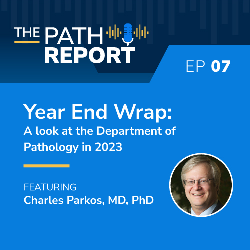
Year End Wrap: A look at the Department of Pathology in 2023
December 19 2023
S1, EP7
Chair of the Department of Pathology, Dr. Charles Parkos, stops by The Path Report to discuss some of the top highlights of 2023 and what he is most excited about in 2024.
The University of Michigan Department of Pathology podcast, The Path Report provides listeners with the inside scoop on the interesting things happening in pathology at Michigan Medicine.
Find it on all the providers below:
![]() Acast
Acast
Amazon Music
![]() Google Podcast
Google Podcast
Spotify
YouTube
More News NEWS ARCHIVE SUBMIT STORY
April 24
Another Step Toward Healthcare Equity: Dr. Jennifer Jones and Colleagues Participate in National Study
April 23
Drs. Sarah Farran and Isabella Holmes selected as Assistant Chief Residents
April 23
Dr. Asma Nusrat Receives 2024 Rous-Whipple Award at ASIP Meeting
April 12
In Memoriam: Dena Ryan, May 16, 1977 – April 4, 2024
It is with profound sadness that we write about the unexpected passing of Dena Ryan, a member of our Pathology Informatics team who managed MediaLab for the Department of Pathology. Dena was born in Grand Rapids, Michigan and attended Lowell High School in Lowell, Michigan. Upon […]
April 12
MCP Student Gabbi Rozumek awarded 2024 Phyllis M. Wise Biomedical Sciences Graduate Student Award for Excellence in Service
The MCP team is proud to share the news that MCP student Gabrielle "Gabbi" Rozumek is the recipient of the 2024 Phyllis M. Wise Biomedical Sciences Graduate Student Award for Excellence in Service.
Gabbi is very passionate about and dedicated to serving her community. Since 2016, […]
April 11
 ON THE COVER
 ON THE COVER
 ON THE COVER
 ON THE COVER
 ON THE COVER
 ON THE COVER
 ON THE COVER
 ON THE COVER
 ON THE COVER