
Where do I look for my scanned slide images?
The interface you will use for viewing slides partially depends upon what you communicate to us through the slide scan request form. Michigan Medicine Pathology faculty, House Officers, and staff should consider the search engine within the "Tumor Boards" application inside the Pathology Clinical Lab Portal as their "go-to" interface for viewing scanned slides. This, of course, does not preclude the use of other viewing interfaces. However, the CLP search engine's ease of use and other features recommend it as the principal resource for digital slide viewing.
Can you put all my slides together in a single folder?
The short answer to this question is "yes." However, it is important to have an understanding of how the folder exists on the server, how its content is accessed, and what it will look like from the end user's experience.
Because Slide Scanning staff can access digital slide content with a direct connection to the server, which end users cannot, we see folders just as you would see them in a standard Windows File Explorer interface. When instructed to create specific folders for specific purposes, we do so. Absent specific instructions, we follow past practices based on the use case indicated to us through the slide scan request form.
The end user accesses digital slide images using one of our interfaces below. The most commonly used interface is the search engine on the Clinical Lab Portal. The CLP search engine lacks the ability to return groups of images organized by folder, however. Our vendor-created database, eSlide Manager, allows a user to return groups of slides organized by folder, but there is a learning curve associated with it. We are happy to teach interested individuals how to make use of the advanced search techniques in eSlide Manager. In the case of the public-facing "IM2," folder icons are visible, discrete, and easily understandable. However, IM2 operates within a set of constraints, as described below.
a. Searches the entirety of our scanned slide collection
b. Can open scanned slides directly using either ImageScope or WebScope
c. Copy of Path Lab report provided if available
d. Behind U-M firewall; access is limited to those Pathology faculty and staff with permission to access the "Tumor Boards" application within the Portal.
e. If a slide has been archived, it will be documented on the interface. If a copy of the image is on the Virtual Slide Box (a server separate from our main one), a link will be provided.
a. Vendor-provided whole-slide image database and image management solution
b. Pros: can execute more sophisticated (Boolean, combinatorial) searches within our available scanned slides. May provide more metadata for a given slide or slide set vs the Clinical Lab Portal search engine.
c. Cons: less user-friendly vs the Clinical Lab Portal search engine; requires more training to learn how to use it. Will not return results from the entirety of our scanned slide library
d. Is behind the UM firewall; access is limited to those faculty and staff with an account.
e. If a slide has been archived, the image cannot be opened, and no thumbnail will be visible. No link to the VSB is provided if a copy of the image is located there.
a. Information and images are publicly available; different server resource than the one housing our principal digital slide library.
b. Intended only for slides with educational purpose
c. Because slides here are publicly accessible, content on the VSB consists of image copies that have been de-identified. A copy of the digital slide with the captured slide label image and any identifying information on it is maintained on our server behind the UM firewall.
d. Indicate your interest in having de-identified slide copies pushed to the VSB by making use of the "radio button" on the slide scan request form. In addition, please notify us in the comments which collection you wish them to be included in.
e. NOTE: as of November, 2020, the VSB features an option for users of opening slides with a local copy of "ImageScope" software or by using "WebScope" to open images within their browser. However, this particular "WebScope" instance is Flash-based, and Flash will be permanently blocked on web browsers after December 31, 2020. Pathology Informatics is working on solutions. Thank you for your patience.
a. Information and images are publicly available; different server resource than the one housing our principal digital slide library.
b. Intended as an alternate resource for, with certain exceptions, small-scale, time-limited, low-frequency slide scans that need to be publicly available. Examples include but are not limited to:
1. Images needed for presentations when a UM faculty member travels
2. Images that need to be shared with professional colleagues outside Michigan Medicine
c. Because slides here are publicly accessible, content on "IM2" consists of image copies that have been de-identified. A copy of the digital slide with the captured slide label image and any identifying information on it is maintained on our server behind the UM firewall.
d. If the IM2 resource seems like it would be a good fit for your digital slide image goals, please contact the Slide Scanning Service first to discuss.
Slides scanned for tumor boards are assigned to a dated web page within the "Tumor Boards" section of the Pathology Clinical Lab Portal. A screenshot from the main interface is shown below. Note: not all conferences shown here.
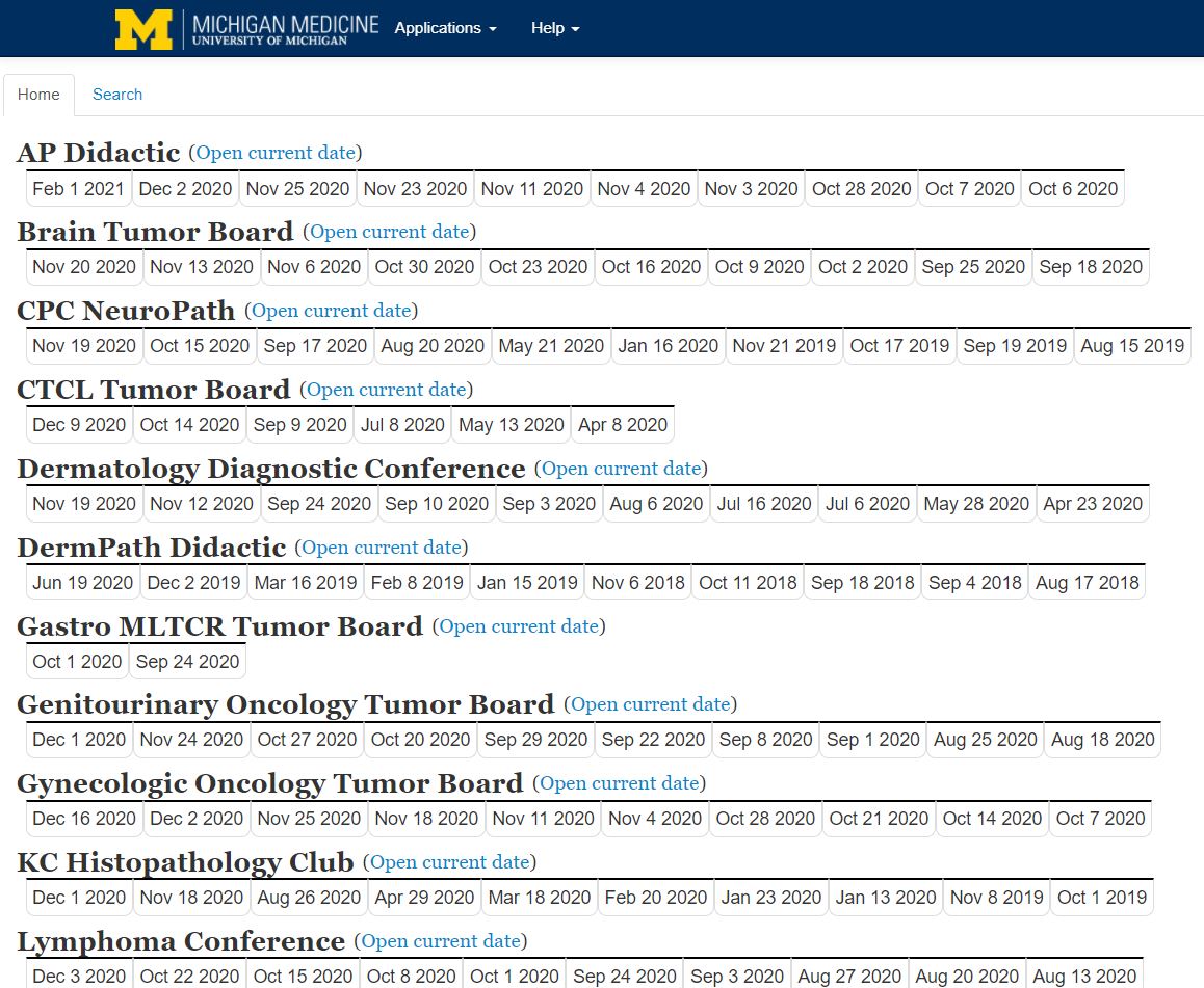
Simply click the appropriate date and a list of cases will appear. Each case will have a link to the Pathology report next to the last name. To open all case images at once, click the last name. To open a single slide, click the stain name.
You may select the modality for displaying the scanned images. "ImageScope" is the vendor's software and must be installed locally on the workstation. "WebScope" will open the images within your web browser.
You will also utilize the "Tumor Boards" interface. When entering the scan request, choose "Didactic" for "Group." You then have several options. Choose the conference name that best fits your purpose.
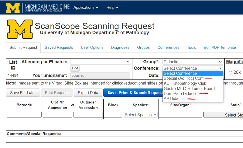
After selecting a Conference name, click in the "Date" field and click the day you wish to designate for the conference. Proceed with filling out the remainder of the form as usual. When the slides are scanned, you will be notified. Look for them under the appropriate conference name within the Tumor Boards section of the Portal (see above). Click the date you requested in the form.
Please contact Digital Pathology directly for these kinds of requests. Slides shared with persons or groups outside U-M will necessarily need to be on a server with a public IP. Therefore they must be de-identified to remove any protected health information (PHI) You may email slidescanningserv [at] med.umich.edu or phone 4-4003.
In the past we have steered our research lab colleagues to the eSlide Manager picture-archive database for viewing their slides. We now recommend to all new research clients that they receive copies of their digital slides that they can save on their own platforms. This is for two reasons - 1) eSlide Manager is less user-friendly than other programs and 2) many researchers are looking to perform image analysis on whole-slide images, which cannot be done through eSM. When you indicate "Research" in the "Group" field, you will be required to indicate via "radio button" icon your preference for viewing your slides. "MBox or Dropbox" is most frequently chosen.
NOTE 1: The scanning process results in ".svs" image files. These image files are archived on a secure server, and copies may be given upon request. Currently 40X scans result in image files that average 0.5 to 1.0 GB each. The file size varies directly with the size of the tissue slice. Image file sizes can range from 0.1 to 3.0 GB. Please make sure you have a storage device of appropriate capacity prior to requesting copies of a group of image files.
NOTE 2: Any SVS files containing protected health information (PHI) can only be transferred through a Dropbox shared account set up to handle sensitive information, or by the use of a HIPAA-compliant encrypted external drive, such as an Apricorn Aegis Padlock.
Clients that elect to receive copies of their svs files can open them using the freeware ImageScope. With ImageScope, clients can extract jpg images suitable for PowerPoint presentations, posters and publications. Multiple images can be compared simultaneously. Areas of interest can be annotated with arrows, shape tools, and labels for downstream presentation or teaching purposes. Finally, as of March, 2022, there is no Mac-compatible version of ImageScope software. An alternative, Mac-compatible product is "QuPath." Link here: https://qupath.github.io/
 ON THE COVER
Breast team reviewing a patient's slide. (From left to right) Ghassan Allo, Fellow; Laura Walters, Clinical Lecturer; Celina Kleer, Professor. See Article |
newsletter
INSIDE PATHOLOGYAbout Our NewsletterInside Pathology is an newsletter published by the Chairman's Office to bring news and updates from inside the department's research and to become familiar with those leading it. It is our hope that those who read it will enjoy hearing about those new and familiar, and perhaps help in furthering our research. CONTENTS
|
 ON THE COVER
Autopsy Technician draws blood while working in the Wayne County morgue. See Article |
newsletter
INSIDE PATHOLOGYAbout Our NewsletterInside Pathology is an newsletter published by the Chairman's Office to bring news and updates from inside the department's research and to become familiar with those leading it. It is our hope that those who read it will enjoy hearing about those new and familiar, and perhaps help in furthering our research. CONTENTS
|
 ON THE COVER
Dr. Sriram Venneti, MD, PhD and Postdoctoral Fellow, Chan Chung, PhD investigate pediatric brain cancer. See Article |
newsletter
INSIDE PATHOLOGYAbout Our NewsletterInside Pathology is an newsletter published by the Chairman's Office to bring news and updates from inside the department's research and to become familiar with those leading it. It is our hope that those who read it will enjoy hearing about those new and familiar, and perhaps help in furthering our research. CONTENTS
|
 ON THE COVER
Director of the Neuropathology Fellowship, Dr. Sandra Camelo-Piragua serves on the Patient and Family Advisory Council. |
newsletter
INSIDE PATHOLOGYAbout Our NewsletterInside Pathology is an newsletter published by the Chairman's Office to bring news and updates from inside the department's research and to become familiar with those leading it. It is our hope that those who read it will enjoy hearing about those new and familiar, and perhaps help in furthering our research. CONTENTS
|
 ON THE COVER
Residents Ashley Bradt (left) and William Perry work at a multi-headed scope in our new facility. |
newsletter
INSIDE PATHOLOGYAbout Our NewsletterInside Pathology is an newsletter published by the Chairman's Office to bring news and updates from inside the department's research and to become familiar with those leading it. It is our hope that those who read it will enjoy hearing about those new and familiar, and perhaps help in furthering our research. CONTENTS
|
 ON THE COVER
Dr. Kristine Konopka (right) instructing residents while using a multi-headed microscope. |
newsletter
INSIDE PATHOLOGYAbout Our NewsletterInside Pathology is an newsletter published by the Chairman's Office to bring news and updates from inside the department's research and to become familiar with those leading it. It is our hope that those who read it will enjoy hearing about those new and familiar, and perhaps help in furthering our research. CONTENTS
|
 ON THE COVER
Patient specimens poised for COVID-19 PCR testing. |
newsletter
INSIDE PATHOLOGYAbout Our NewsletterInside Pathology is an newsletter published by the Chairman's Office to bring news and updates from inside the department's research and to become familiar with those leading it. It is our hope that those who read it will enjoy hearing about those new and familiar, and perhaps help in furthering our research. CONTENTS
|
 ON THE COVER
Dr. Pantanowitz demonstrates using machine learning in analyzing slides. |
newsletter
INSIDE PATHOLOGYAbout Our NewsletterInside Pathology is an newsletter published by the Chairman's Office to bring news and updates from inside the department's research and to become familiar with those leading it. It is our hope that those who read it will enjoy hearing about those new and familiar, and perhaps help in furthering our research. CONTENTS
|
 ON THE COVER
(Left to Right) Drs. Angela Wu, Laura Lamps, and Maria Westerhoff. |
newsletter
INSIDE PATHOLOGYAbout Our NewsletterInside Pathology is an newsletter published by the Chairman's Office to bring news and updates from inside the department's research and to become familiar with those leading it. It is our hope that those who read it will enjoy hearing about those new and familiar, and perhaps help in furthering our research. CONTENTS
|
 ON THE COVER
Illustration representing the various machines and processing used within our labs. |
newsletter
INSIDE PATHOLOGYAbout Our NewsletterInside Pathology is an newsletter published by the Chairman's Office to bring news and updates from inside the department's research and to become familiar with those leading it. It is our hope that those who read it will enjoy hearing about those new and familiar, and perhaps help in furthering our research. CONTENTS
|
 ON THE COVER
Rendering of the D. Dan and Betty Khn Health Care Pavilion. Credit: HOK |
newsletter
INSIDE PATHOLOGYAbout Our NewsletterInside Pathology is an newsletter published by the Chairman's Office to bring news and updates from inside the department's research and to become familiar with those leading it. It is our hope that those who read it will enjoy hearing about those new and familiar, and perhaps help in furthering our research. CONTENTS
|
MLabs, established in 1985, functions as a portal to provide pathologists, hospitals. and other reference laboratories access to the faculty, staff and laboratories of the University of Michigan Health System’s Department of Pathology. MLabs is a recognized leader for advanced molecular diagnostic testing, helpful consultants and exceptional customer service.